
Search Count: 52
 |
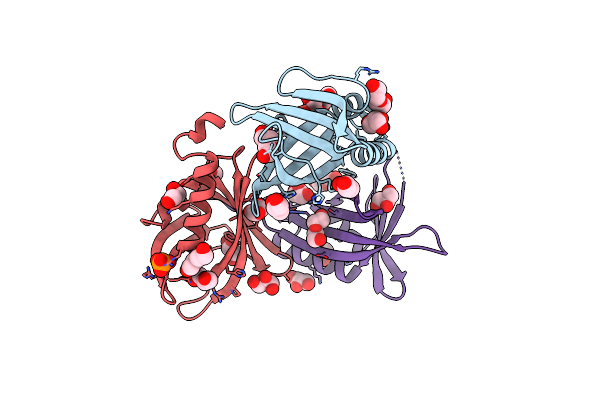
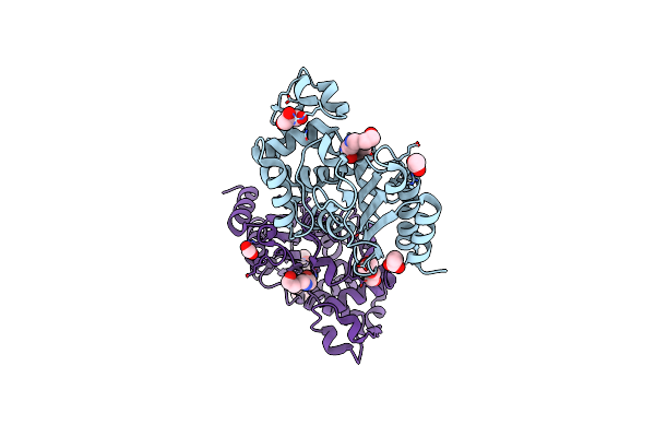
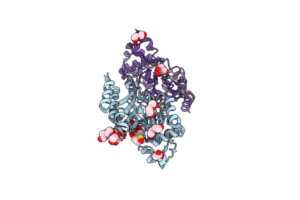
N-Acetylglucosamine 6-Phosphate Dehydratase: Inhibited 6-Phosphogluconic Acid State Of Nags
Organism: Streptomyces coelicolor
Method: X-RAY DIFFRACTION Resolution:1.69 Å Release Date: 2025-02-19 Classification: SUGAR BINDING PROTEIN Ligands: SO4, 6PG |
 |
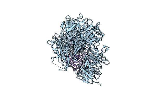
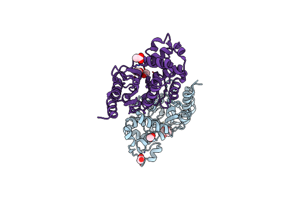
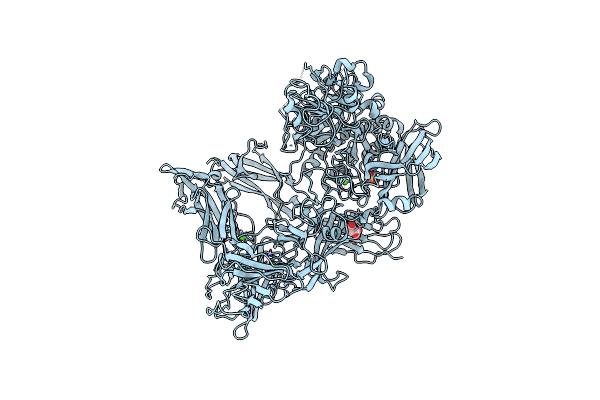
Organism: Streptomyces coelicolor
Method: X-RAY DIFFRACTION Resolution:2.30 Å Release Date: 2025-02-19 Classification: SUGAR BINDING PROTEIN Ligands: SO4 |
 |
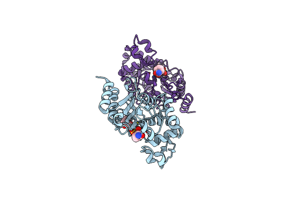
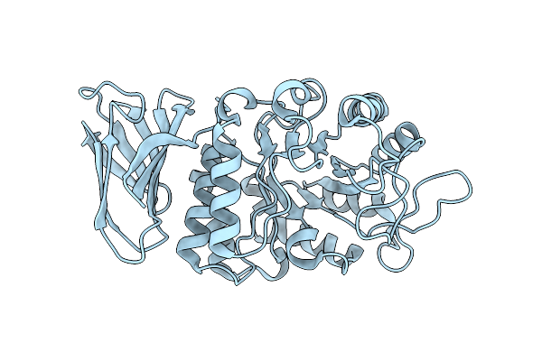
N-Acetylglucosamine 6-Phosphate Dehydratase: Glcnac6P Substrate-Bound State Of Nags
Organism: Streptomyces coelicolor
Method: X-RAY DIFFRACTION Resolution:2.59 Å Release Date: 2025-02-19 Classification: SUGAR BINDING PROTEIN Ligands: SO4, 16G |
 |
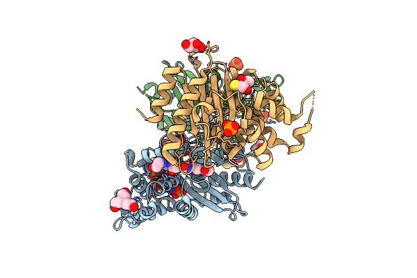
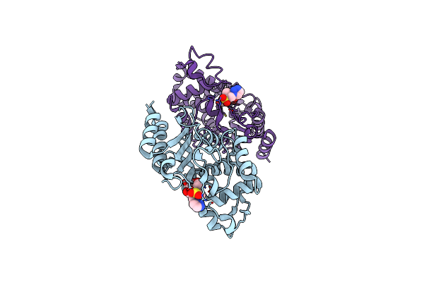
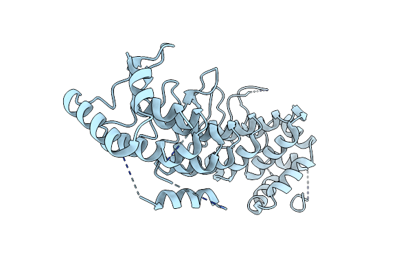
Mutations In Ssgb Correlate To Longitudinal Cell Division During Sporulation Of Streptomyces Coelicolor
Organism: Streptomyces sp. ag82_o1-9
Method: X-RAY DIFFRACTION Resolution:2.30 Å Release Date: 2020-08-26 Classification: CELL CYCLE Ligands: PEG, GOL, PO4 |
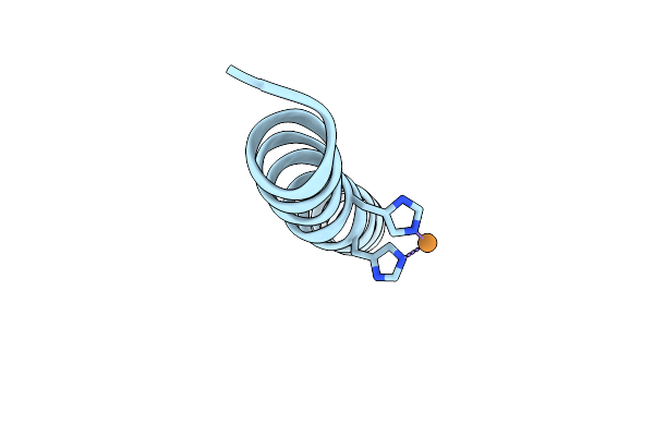 |
Selective Formation Of Trinuclear Transition Metal Centers In A Trimeric Helical Peptide
Organism: Synthetic construct
Method: X-RAY DIFFRACTION Resolution:2.35 Å Release Date: 2019-09-04 Classification: DE NOVO PROTEIN Ligands: CU |
 |
Organism: Homo sapiens
Method: X-RAY DIFFRACTION Resolution:2.92 Å Release Date: 2019-01-30 Classification: PROTEIN BINDING |
 |
Organism: Mycobacterium tuberculosis
Method: X-RAY DIFFRACTION Resolution:1.63 Å Release Date: 2019-01-23 Classification: HYDROLASE Ligands: EDO, PG0 |
 |
Structure Of S70A Blac From Mycobacterium Tuberculosis Obtained From Crystals Produced In The Absence Of Dtt
Organism: Mycobacterium tuberculosis
Method: X-RAY DIFFRACTION Resolution:1.19 Å Release Date: 2019-01-23 Classification: HYDROLASE Ligands: 15P, PEG, GOL |
 |
Structure Of S70A Blac From Mycobacterium Tuberculosis Obtained From Crystals Produced In The Presence Of Dtt
Organism: Mycobacterium tuberculosis
Method: X-RAY DIFFRACTION Resolution:2.54 Å Release Date: 2019-01-23 Classification: HYDROLASE Ligands: TAM, DTT, PO4, GOL |
 |
Structure Of Blac From Mycobacterium Tuberculosis Bound To The Trans-Enamine Adduct Derived From Clavulanic Acid.
Organism: Mycobacterium tuberculosis
Method: X-RAY DIFFRACTION Resolution:1.93 Å Release Date: 2019-01-23 Classification: HYDROLASE Ligands: ISS, ACT, GOL, 15P, CO2 |
 |
Structure Of Blac From Mycobacterium Tuberculosis Bound To The Propionaldehyde Ester Adduct Of Clavulanic Acid.
Organism: Mycobacterium tuberculosis
Method: X-RAY DIFFRACTION Release Date: 2019-01-23 Classification: HYDROLASE Ligands: FK2, ACT |
 |
Structure Of Blac From Mycobacterium Tuberculosis Covalently Bound To Avibactam.
Organism: Mycobacterium tuberculosis
Method: X-RAY DIFFRACTION Resolution:1.62 Å Release Date: 2019-01-23 Classification: HYDROLASE Ligands: NXL, 15P |
 |
Structure Of Blac From Mycobacterium Tuberculosis Bound To The Trans-Enamine Adduct Of Tazobactam.
Organism: Mycobacterium tuberculosis
Method: X-RAY DIFFRACTION Resolution:2.72 Å Release Date: 2019-01-23 Classification: HYDROLASE Ligands: TBE, ACT |
 |
Structure Of Blac From Mycobacterium Tuberculosis Bound To The Trans-Enamine Adduct Of Sulbactam.
Organism: Mycobacterium tuberculosis
Method: X-RAY DIFFRACTION Resolution:1.90 Å Release Date: 2019-01-23 Classification: HYDROLASE Ligands: TSL, PEG, GOL, ACT |
 |
Organism: Streptococcus pyogenes
Method: X-RAY DIFFRACTION Resolution:3.09 Å Release Date: 2018-08-08 Classification: LYASE Ligands: SO4, CA |
 |
Organism: Streptococcus pyogenes
Method: X-RAY DIFFRACTION Resolution:3.00 Å Release Date: 2018-08-08 Classification: LYASE Ligands: AES, SO4, CA |
 |
Organism: Streptococcus pyogenes
Method: X-RAY DIFFRACTION Resolution:2.80 Å Release Date: 2018-08-08 Classification: LYASE Ligands: CL, CA, SO4, MLA |
 |
Organism: Nicotiana benthamiana
Method: X-RAY DIFFRACTION Resolution:2.80 Å Release Date: 2018-04-25 Classification: HYDROLASE |
 |
Organism: Legionella pneumophila subsp. pneumophila (strain philadelphia 1 / atcc 33152 / dsm 7513)
Method: X-RAY DIFFRACTION Resolution:3.21 Å Release Date: 2017-11-29 Classification: PROTEIN BINDING |
 |
Crystal Structure Of Blac From Mycobacterium Tuberculosis Bound To Phosphate
Organism: Mycobacterium tuberculosis
Method: X-RAY DIFFRACTION Resolution:1.19 Å Release Date: 2017-11-15 Classification: HYDROLASE Ligands: PO4, ACT, ETE, AE4 |