
Search Count: 39
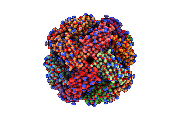 |
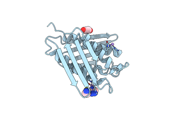
Imidazole Glycerol Phosphate Dehydratase From Mycobacterium Tuberculosis, Apo Structure
Organism: Mycobacterium tuberculosis
Method: ELECTRON MICROSCOPY Release Date: 2025-06-18 Classification: LYASE Ligands: MN |
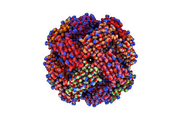 |
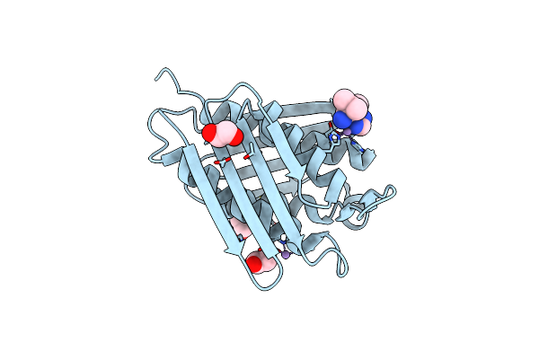
Imidazole Glycerol Phosphate Dehydratase From Mycobacterium Tuberculosis, In Complex With Aminotriazole
Organism: Mycobacterium tuberculosis
Method: ELECTRON MICROSCOPY Release Date: 2025-06-18 Classification: LYASE Ligands: MN, 3TR |
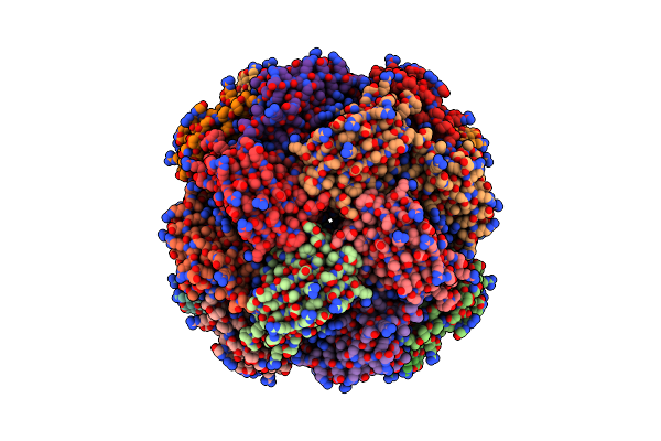 |
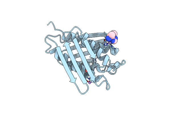
Imidazole Glycerol Phosphate Dehydratase From Mycobacterium Tuberculosis, Apo Structure
Organism: Mycobacterium tuberculosis
Method: ELECTRON MICROSCOPY Release Date: 2025-06-18 Classification: LYASE Ligands: MN |
 |
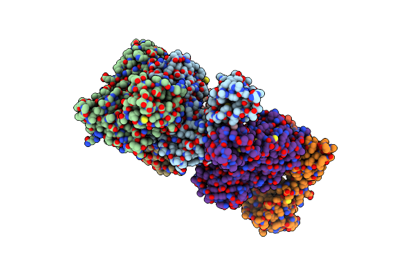
Organism: Staphylococcus aureus subsp. aureus col
Method: X-RAY DIFFRACTION Resolution:2.30 Å Release Date: 2025-02-12 Classification: TRANSFERASE Ligands: ZN, SO4, CL |
 |
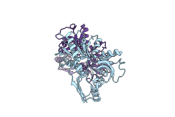
Crystal Structure Of Rab5B Gtpase Domain From Leishmania Donovani In Complex With Gdp
Organism: Leishmania donovani
Method: X-RAY DIFFRACTION Resolution:2.50 Å Release Date: 2025-01-15 Classification: ENDOCYTOSIS Ligands: MG, GDP |
 |
Organism: Mycobacterium tuberculosis
Method: X-RAY DIFFRACTION Resolution:1.75 Å Release Date: 2024-10-09 Classification: LYASE Ligands: MN, CL, PEG, EDO |
 |
Organism: Mycobacterium tuberculosis h37rv
Method: X-RAY DIFFRACTION Resolution:3.15 Å Release Date: 2024-07-03 Classification: DNA BINDING PROTEIN |
 |
Crystal Structure Of Gppnhp Bound Gtpase Domain Of Rab5A From Leishmania Donovani
Organism: Leishmania donovani
Method: X-RAY DIFFRACTION Resolution:1.80 Å Release Date: 2024-06-19 Classification: ENDOCYTOSIS Ligands: EDO, GNP, MG |
 |
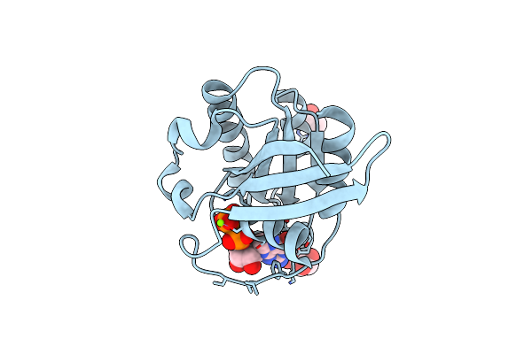
Crystal Structure Of Peptidyl-Trna Hydrolase Mutant From Enterococcus Faecium
Organism: Enterococcus faecium
Method: X-RAY DIFFRACTION Resolution:2.90 Å Release Date: 2024-04-03 Classification: HYDROLASE Ligands: GOL |
 |
Crystal Structure Of Padr- Family Transcriptional Regulator Rv1176C From Mycobacterium Tuberculosis H37Rv.
Organism: Mycobacterium tuberculosis h37rv
Method: X-RAY DIFFRACTION Resolution:2.94 Å Release Date: 2024-03-13 Classification: DNA BINDING PROTEIN Ligands: GOL, CL, P6G |
 |
Organism: Enterococcus faecium
Method: X-RAY DIFFRACTION Resolution:1.92 Å Release Date: 2024-01-17 Classification: HYDROLASE |
 |
Organism: Pyrococcus horikoshii ot3
Method: X-RAY DIFFRACTION Resolution:2.00 Å Release Date: 2022-07-20 Classification: TRANSCRIPTION Ligands: ILE, EDO |
 |
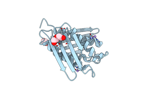
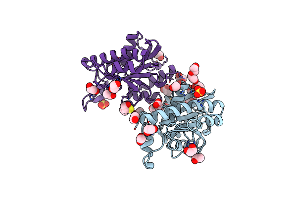
Crystal Structure Of M.Tuberculosis Imidazole Glycerol Phosphate Dehydratase In Complex With An Inhibitor
Organism: Mycobacterium tuberculosis (strain atcc 25618 / h37rv)
Method: X-RAY DIFFRACTION Resolution:1.85 Å Release Date: 2022-07-20 Classification: LYASE Ligands: MN, 47Z, EDO, CL, ACT |
 |
Crystal Structure Of The Padr-Family Transcriptional Regulator Rv3488 Of Mycobacterium Tuberculosis H37Rv In Complex With Manganese Ion
Organism: Mycobacterium tuberculosis h37rv
Method: X-RAY DIFFRACTION Resolution:2.80 Å Release Date: 2022-02-09 Classification: DNA BINDING PROTEIN Ligands: MN |
 |
Crystal Structure Of M.Tuberculosis Imidazole Glycerol Phosphate Dehydratase In Complex With An Inhibitor
Organism: Mycobacterium tuberculosis (strain atcc 25618 / h37rv)
Method: X-RAY DIFFRACTION Resolution:2.28 Å Release Date: 2021-12-22 Classification: LYASE Ligands: MN, CL, HB3, EDO |
 |
Crystal Structure Of M.Tuberculosis Imidazole Glycerol Phosphate Dehydratase In Complex With An Inhibitor
Organism: Mycobacterium tuberculosis (strain atcc 25618 / h37rv)
Method: X-RAY DIFFRACTION Resolution:2.20 Å Release Date: 2021-11-10 Classification: LYASE/LYASE INHIBITOR Ligands: MN, H3L, GOL, CL |
 |
Adenosine Triphosphate Phosphoribosyltransferase From Vibrio Cholerae In Complex With Atp And Prpp
Organism: Vibrio cholerae serotype o1 (strain m66-2)
Method: X-RAY DIFFRACTION Resolution:2.92 Å Release Date: 2021-10-20 Classification: TRANSFERASE Ligands: PEG, EDO, PO4, GOL, MG, ATP, PRP |
 |
Organism: Vibrio cholerae serotype o1 (strain m66-2)
Method: X-RAY DIFFRACTION Resolution:2.70 Å Release Date: 2021-10-20 Classification: TRANSFERASE |
 |
Organism: Klebsiella pneumoniae
Method: X-RAY DIFFRACTION Resolution:1.89 Å Release Date: 2021-03-17 Classification: HYDROLASE Ligands: PGE, PEG, EDO, ACT, CL, SO4, BME |
 |
Crystal Structure Of Stabilized Rab5A Gtpase Domain From Leishmania Donovani
Organism: Leishmania donovani
Method: X-RAY DIFFRACTION Resolution:1.80 Å Release Date: 2020-11-11 Classification: ENDOCYTOSIS Ligands: GDP, GOL, ACT, MG |