
Search Count: 48
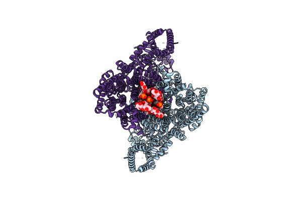 |
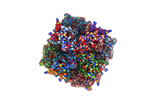
Organism: Strongylocentrotus purpuratus
Method: ELECTRON MICROSCOPY Release Date: 2025-03-26 Classification: MEMBRANE PROTEIN Ligands: POV, AJP |
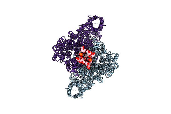 |
Organism: Strongylocentrotus purpuratus
Method: ELECTRON MICROSCOPY Release Date: 2025-03-26 Classification: MEMBRANE PROTEIN Ligands: POV, AJP |
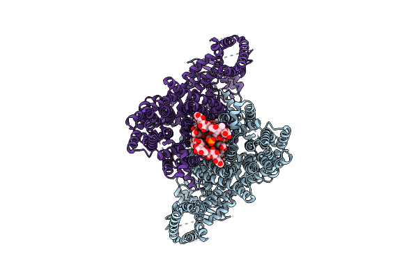 |
Organism: Strongylocentrotus purpuratus
Method: ELECTRON MICROSCOPY Release Date: 2025-03-26 Classification: MEMBRANE PROTEIN Ligands: POV, AJP |
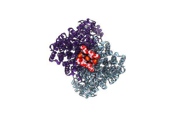 |
Organism: Strongylocentrotus purpuratus
Method: ELECTRON MICROSCOPY Release Date: 2025-03-26 Classification: MEMBRANE PROTEIN Ligands: POV, AJP |
 |
Organism: Strongylocentrotus purpuratus
Method: ELECTRON MICROSCOPY Release Date: 2025-03-26 Classification: MEMBRANE PROTEIN Ligands: POV, AJP |
 |
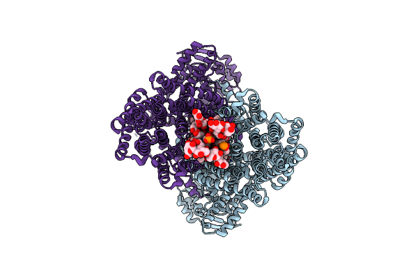
Organism: Strongylocentrotus purpuratus
Method: ELECTRON MICROSCOPY Release Date: 2025-03-26 Classification: MEMBRANE PROTEIN Ligands: POV, AJP, CMP |
 |
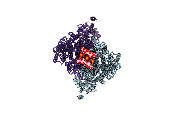
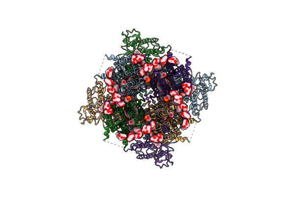
Wt Sea Urchin Slc9C1 With 5Mm Camp At Ph 6 In Na+ - Grip And Twist (Gnt) Conformation
Organism: Strongylocentrotus purpuratus
Method: ELECTRON MICROSCOPY Release Date: 2025-03-26 Classification: MEMBRANE PROTEIN Ligands: POV, AJP, CMP |
 |
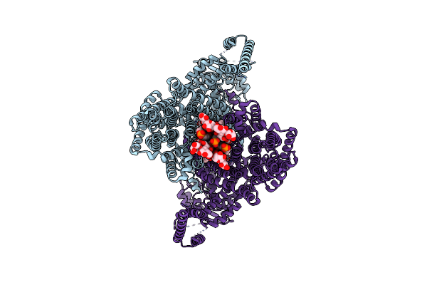
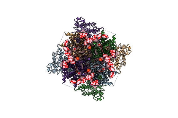
Wt Sea Urchin Slc9C1 With 5Mm Camp At Ph 6 In Na+ - Grip And Twist Like (Gntl) Conformation
Organism: Strongylocentrotus purpuratus
Method: ELECTRON MICROSCOPY Release Date: 2025-03-26 Classification: MEMBRANE PROTEIN Ligands: POV, AJP, CMP |
 |
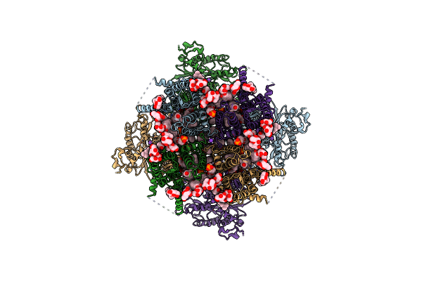
Organism: Strongylocentrotus purpuratus
Method: ELECTRON MICROSCOPY Release Date: 2025-03-26 Classification: MEMBRANE PROTEIN Ligands: POV, AJP |
 |
Organism: Strongylocentrotus purpuratus
Method: ELECTRON MICROSCOPY Release Date: 2025-03-26 Classification: MEMBRANE PROTEIN Ligands: POV, AJP |
 |
Organism: Homo sapiens
Method: ELECTRON MICROSCOPY Release Date: 2024-12-25 Classification: TRANSPORT PROTEIN Ligands: POV, K, AJP, CLR, NAG |
 |
Organism: Homo sapiens
Method: ELECTRON MICROSCOPY Release Date: 2024-12-25 Classification: TRANSPORT PROTEIN Ligands: K |
 |
Organism: Homo sapiens
Method: ELECTRON MICROSCOPY Release Date: 2024-12-11 Classification: TRANSPORT PROTEIN Ligands: K, POV, CLR, AJP |
 |
Organism: Homo sapiens
Method: ELECTRON MICROSCOPY Release Date: 2024-12-11 Classification: TRANSPORT PROTEIN Ligands: K, POV, CLR, AJP |
 |
Organism: Homo sapiens
Method: ELECTRON MICROSCOPY Release Date: 2024-12-11 Classification: TRANSPORT PROTEIN Ligands: K, POV, CLR, AJP |
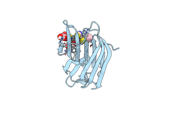 |
Organism: Homo sapiens
Method: X-RAY DIFFRACTION Resolution:1.14 Å Release Date: 2023-11-08 Classification: SUGAR BINDING PROTEIN Ligands: Y58, SCN |
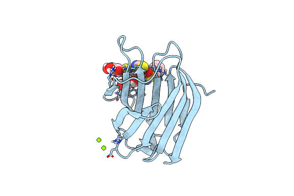 |
Organism: Homo sapiens
Method: X-RAY DIFFRACTION Resolution:1.09 Å Release Date: 2023-11-08 Classification: SUGAR BINDING PROTEIN Ligands: SCN, YJO, MG |
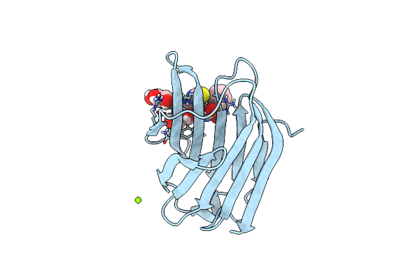 |
Organism: Homo sapiens
Method: X-RAY DIFFRACTION Resolution:1.08 Å Release Date: 2023-11-08 Classification: SUGAR BINDING PROTEIN Ligands: SCN, YJF, MG |
 |
Organism: Homo sapiens
Method: X-RAY DIFFRACTION Resolution:2.20 Å Release Date: 2022-04-13 Classification: SIGNALING PROTEIN Ligands: EPE, GOL |
 |
Crystal Structure Of A Standalone Versatile Eal Protein From Vibrio Cholerae O395 - C-Di-Imp Bound Form
Organism: Vibrio cholerae serotype o1 (strain atcc 39541 / classical ogawa 395 / o395)
Method: X-RAY DIFFRACTION Resolution:2.20 Å Release Date: 2020-06-17 Classification: HYDROLASE Ligands: C2I, CA |