
Search Count: 27
 |
Organism: Enterobacter cloacae
Method: X-RAY DIFFRACTION Resolution:2.30 Å Release Date: 2018-10-31 Classification: TRANSPORT PROTEIN Ligands: SO4, BOG, C8E |
 |
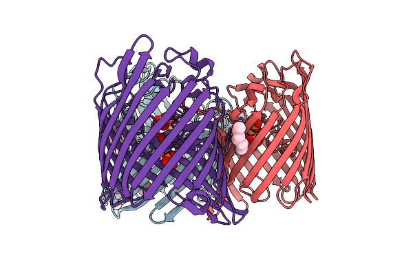
Organism: Klebsiella pneumoniae
Method: X-RAY DIFFRACTION Resolution:1.50 Å Release Date: 2018-06-20 Classification: MEMBRANE PROTEIN Ligands: TRS, C8E |
 |
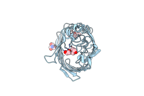
Organism: Klebsiella pneumoniae
Method: X-RAY DIFFRACTION Resolution:1.65 Å Release Date: 2018-06-20 Classification: MEMBRANE PROTEIN Ligands: C8E, MG |
 |
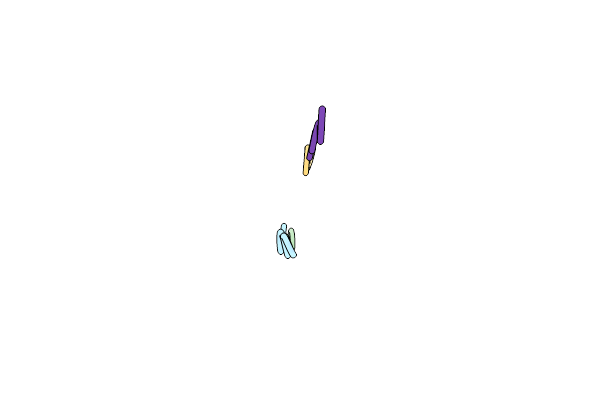
Structure Of 283-Lgny-286, The Steric Zipper That Supports The Self-Association Of P. Stuartii Omp-Pst2 Into Dimers Of Trimers
Organism: Providencia stuartii
Method: X-RAY DIFFRACTION Resolution:1.00 Å Release Date: 2018-02-21 Classification: CELL ADHESION Ligands: SO4 |
 |
Structure Of 206-Gvvtse-211, The Steric Zipper That Supports The Self-Association Of P. Stuartii Omp-Pst1 Into Dimers Of Trimers
Organism: Providencia stuartii
Method: X-RAY DIFFRACTION Resolution:1.91 Å Release Date: 2018-02-21 Classification: CELL ADHESION |
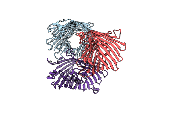 |
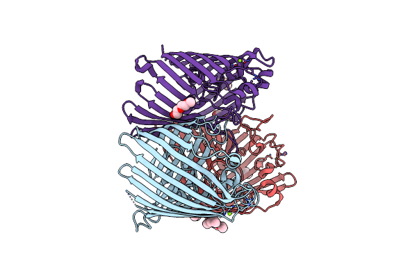
Organism: Providencia stuartii
Method: X-RAY DIFFRACTION Resolution:3.12 Å Release Date: 2018-02-21 Classification: TRANSPORT PROTEIN Ligands: CA |
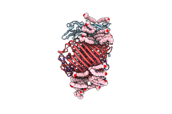 |
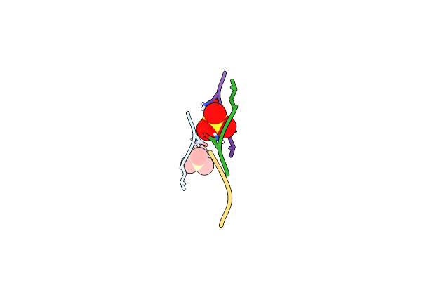
Organism: Providencia stuartii
Method: X-RAY DIFFRACTION Resolution:2.70 Å Release Date: 2018-02-21 Classification: CELL ADHESION Ligands: LDA, CA, CL |
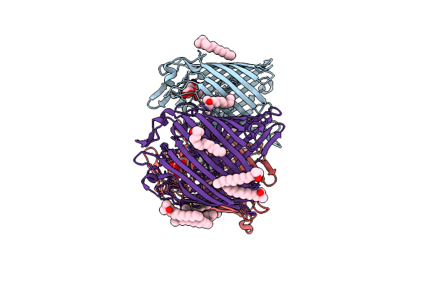 |
Organism: Providencia stuartii
Method: X-RAY DIFFRACTION Resolution:3.00 Å Release Date: 2018-02-21 Classification: CELL ADHESION Ligands: LDA, CA |
 |
Crystal Structure Of Type 2 Pdf From Streptococcus Agalactiae, Crystallized In Imidazole Buffer
Organism: Streptococcus agalactiae
Method: X-RAY DIFFRACTION Resolution:2.00 Å Release Date: 2016-11-30 Classification: HYDROLASE Ligands: IMD, ZN |
 |
Crystal Structure Of Type 2 Pdf From Streptococcus Agalactiae, Crystallized In Cacodylate Buffer
Organism: Streptococcus agalactiae
Method: X-RAY DIFFRACTION Resolution:2.80 Å Release Date: 2016-11-30 Classification: HYDROLASE Ligands: NI |
 |
Crystal Structure Of Type 2 Pdf From Streptococcus Agalactiae In Complex With Tripeptide Met-Ala-Ser
Organism: Streptococcus agalactiae, Escherichia coli
Method: X-RAY DIFFRACTION Resolution:1.70 Å Release Date: 2016-11-30 Classification: HYDROLASE Ligands: ACT, ZN |
 |
Crystal Structure Of Type 2 Pdf From Streptococcus Agalactiae In Complex With Tripeptide Met-Ala-Arg
Organism: Streptococcus agalactiae, Escherichia coli
Method: X-RAY DIFFRACTION Resolution:1.60 Å Release Date: 2016-11-30 Classification: HYDROLASE Ligands: ACT, NI |
 |
Crystal Structure Of Type 2 Pdf From Streptococcus Agalactiae In Complex With Actinonin
Organism: Streptococcus agalactiae
Method: X-RAY DIFFRACTION Resolution:2.00 Å Release Date: 2016-11-30 Classification: HYDROLASE Ligands: BB2, ACT, ZN |
 |
Crystal Structure Of Type 2 Pdf From Streptococcus Agalactiae In Complex With Inhibitor At002
Organism: Streptococcus agalactiae
Method: X-RAY DIFFRACTION Resolution:2.00 Å Release Date: 2016-11-30 Classification: HYDROLASE Ligands: SF7, ACT, IMD, ZN |
 |
Crystal Structure Of Type 2 Pdf From Streptococcus Agalactiae In Complex With Inhibitor At018
Organism: Streptococcus agalactiae
Method: X-RAY DIFFRACTION Resolution:1.60 Å Release Date: 2016-11-30 Classification: HYDROLASE Ligands: SF5, ACT, IMD, ZN |
 |
Crystal Structure Of Type 2 Pdf From Streptococcus Agalactiae In Complex With Inhibitor At019
Organism: Streptococcus agalactiae
Method: X-RAY DIFFRACTION Resolution:2.40 Å Release Date: 2016-11-30 Classification: HYDROLASE Ligands: 6JT, ACT, IMD, ZN |
 |
Crystal Structure Of Type 2 Pdf From Streptococcus Agalactiae In Complex With Inhibitor At020
Organism: Streptococcus agalactiae
Method: X-RAY DIFFRACTION Resolution:1.80 Å Release Date: 2016-11-30 Classification: HYDROLASE Ligands: 7JT, ACT, IMD, ZN |
 |
Crystal Structure Of Type 2 Pdf From Streptococcus Agalactiae In Complex With Inhibitor 6B (Ab47)
Organism: Streptococcus agalactiae
Method: X-RAY DIFFRACTION Resolution:1.70 Å Release Date: 2016-11-30 Classification: HYDROLASE Ligands: BB4, ACT, ZN |
 |
Crystal Structure Of Type 2 Pdf From Streptococcus Agalactiae In Complex With Inhibitor Smp289
Organism: Streptococcus agalactiae
Method: X-RAY DIFFRACTION Resolution:2.10 Å Release Date: 2016-11-30 Classification: HYDROLASE Ligands: 6JU, ACT, IMD, ZN |
 |
Crystal Structure Of Type 2 Pdf From Streptococcus Agalactiae In Complex With Inhibitor Ras358 (21)
Organism: Streptococcus agalactiae
Method: X-RAY DIFFRACTION Resolution:1.80 Å Release Date: 2016-11-30 Classification: HYDROLASE Ligands: PN3, ACT, IMD, ZN |