
Search Count: 29
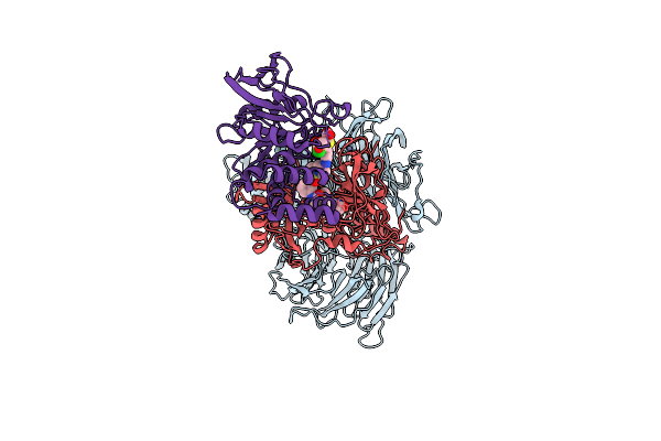 |
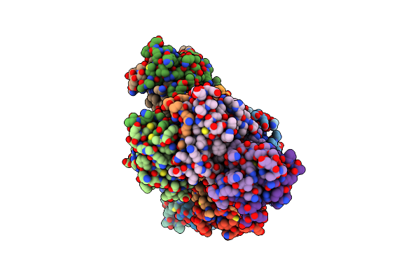
Organism: Homo sapiens
Method: ELECTRON MICROSCOPY Release Date: 2024-09-11 Classification: HYDROLASE Ligands: WO8, ZN |
 |
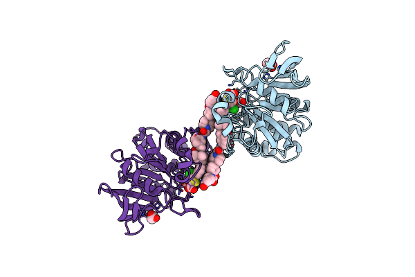
Organism: Mus musculus
Method: ELECTRON MICROSCOPY Release Date: 2024-09-11 Classification: SIGNALING PROTEIN |
 |
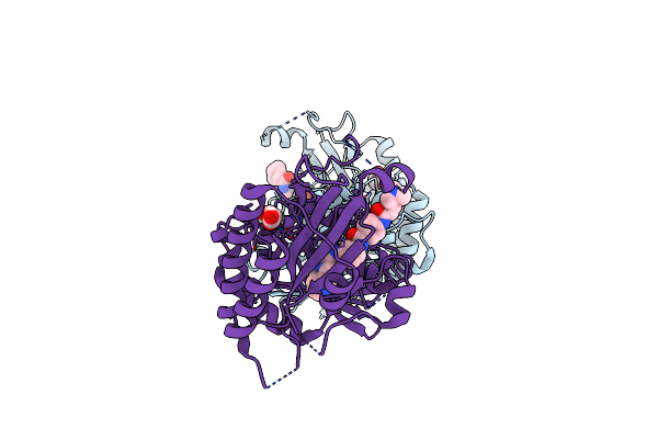
Organism: Homo sapiens
Method: X-RAY DIFFRACTION Resolution:1.93 Å Release Date: 2024-09-04 Classification: HYDROLASE Ligands: UB0, ACT, NA |
 |
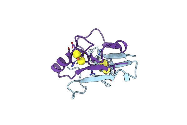
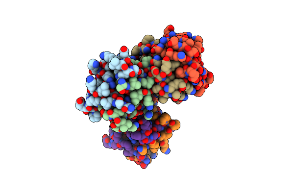
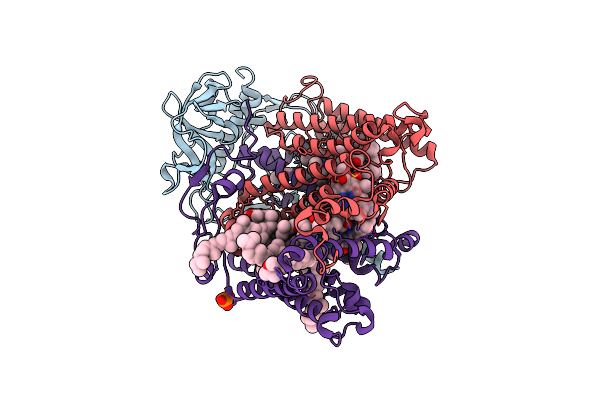
Crystal Structure Of Human T1061E Pi3Kalpha In Complex With Its Regulatory Subunit And The Inhibitor Gdc-0077 (Inavolisib)
Organism: Homo sapiens
Method: X-RAY DIFFRACTION Resolution:2.82 Å Release Date: 2023-12-13 Classification: TRANSFERASE Ligands: MWF, EDO, PGE, CL |
 |
Organism: Homo sapiens
Method: X-RAY DIFFRACTION Resolution:2.04 Å Release Date: 2023-07-26 Classification: SIGNALING PROTEIN/INHIBITOR Ligands: M5R |
 |
Organism: Homo sapiens
Method: X-RAY DIFFRACTION Resolution:2.21 Å Release Date: 2020-04-29 Classification: TRANSFERASE Ligands: N51, GOL |
 |
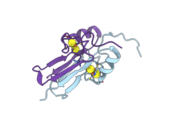
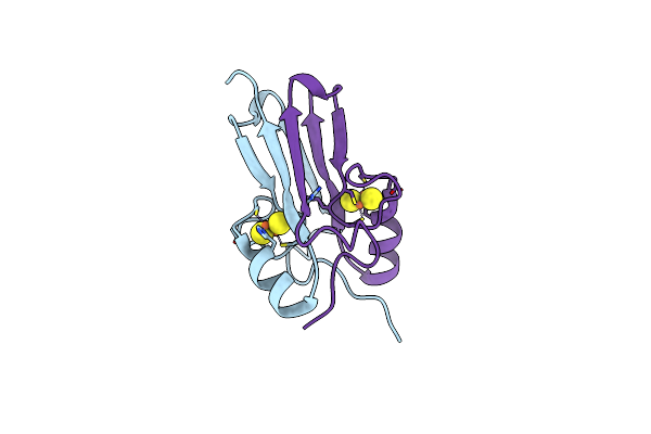
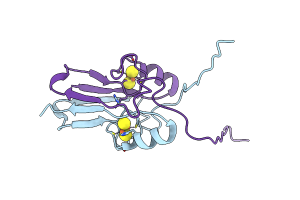
The 1.55A Crystal Structure Of Naf1 (Miner1): The Redox-Active 2Fe-2S Protein
Organism: Homo sapiens
Method: X-RAY DIFFRACTION Resolution:1.65 Å Release Date: 2014-07-02 Classification: METAL BINDING PROTEIN Ligands: FES |
 |
Organism: Homo sapiens
Method: X-RAY DIFFRACTION Resolution:1.58 Å Release Date: 2014-07-02 Classification: METAL BINDING PROTEIN Ligands: FES |
 |
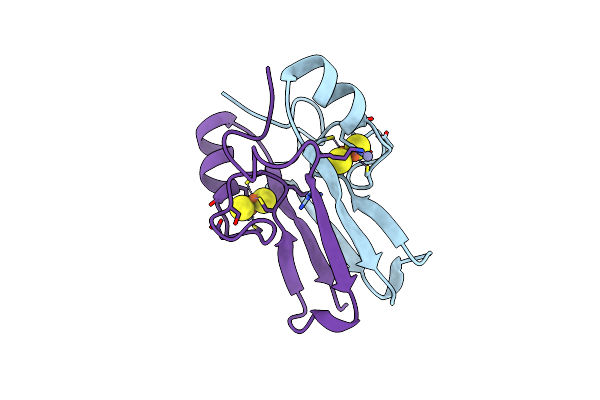
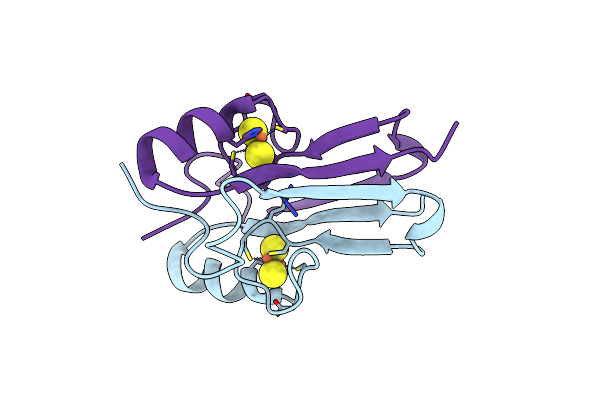
The Crystal Structure Of A Human Mitoneet Mutant With An Ala Inserted Between Asp 67 And Lys 68
Organism: Homo sapiens
Method: X-RAY DIFFRACTION Resolution:1.19 Å Release Date: 2012-12-26 Classification: METAL BINDING PROTEIN Ligands: FES |
 |
The Crystal Structure Of A Human Mitoneet Mutant With Asp 67 Replaced By A Gly
Organism: Homo sapiens
Method: X-RAY DIFFRACTION Resolution:2.40 Å Release Date: 2012-12-26 Classification: METAL BINDING PROTEIN Ligands: FES |
 |
The Crystal Structure Of A Human Mitoneet Mutant With Met 62 Replaced By A Gly
Organism: Homo sapiens
Method: X-RAY DIFFRACTION Resolution:1.55 Å Release Date: 2012-12-26 Classification: METAL BINDING PROTEIN Ligands: FES |
 |
The Crystal Structure Of A Human Mitoneet Double Mutant In Which Gly 66 Are Asp 67 Are Both Replaced With Ala Residues
Organism: Homo sapiens
Method: X-RAY DIFFRACTION Resolution:1.35 Å Release Date: 2012-12-26 Classification: METAL BINDING PROTEIN Ligands: FES |
 |
Organism: Arabidopsis thaliana
Method: X-RAY DIFFRACTION Resolution:1.75 Å Release Date: 2012-05-23 Classification: METAL BINDING PROTEIN Ligands: FES, ZN |
 |
Organism: Arabidopsis thaliana
Method: X-RAY DIFFRACTION Resolution:1.14 Å Release Date: 2012-05-23 Classification: METAL BINDING PROTEIN Ligands: FES |
 |
Organism: Homo sapiens
Method: X-RAY DIFFRACTION Resolution:1.70 Å Release Date: 2011-06-01 Classification: METAL BINDING PROTEIN Ligands: FES |
 |
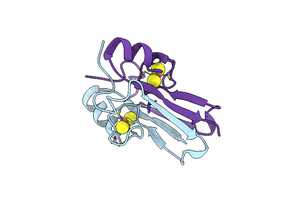
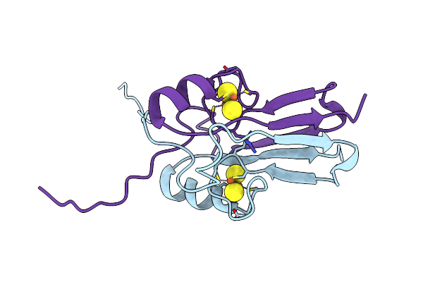
Crystal Structure Of Miner1: The Redox-Active 2Fe-2S Protein Causative In Wolfram Syndrome 2
Organism: Homo sapiens
Method: X-RAY DIFFRACTION Resolution:2.10 Å Release Date: 2009-08-18 Classification: METAL BINDING PROTEIN Ligands: FES |
 |
The Novel 2Fe-2S Outer Mitochondrial Protein Mitoneet Displays Conformational Flexibility In Its N-Terminal Cytoplasmic Tethering Domain
Organism: Homo sapiens
Method: X-RAY DIFFRACTION Resolution:1.40 Å Release Date: 2009-07-07 Classification: METAL BINDING PROTEIN Ligands: FES |
 |
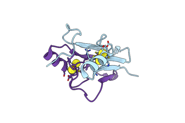
Mitoneet Is A Uniquely Folded 2Fe-2S Outer Mitochondrial Membrane Protein Stabilized By Pioglitazone
Organism: Homo sapiens
Method: X-RAY DIFFRACTION Resolution:1.50 Å Release Date: 2007-08-21 Classification: METAL BINDING PROTEIN Ligands: FES |
 |
Structure Of A Quintuple Mutant Of Photosynthetic Reaction Center From Rhodobacter Sphaeroides
Organism: Rhodobacter sphaeroides
Method: X-RAY DIFFRACTION Resolution:2.25 Å Release Date: 2005-05-17 Classification: PHOTOSYNTHESIS Ligands: BCL, BPH, U10, HTO, LDA, GOL, FE2, CL, PO4, SPO, CDL |
 |
Photosynthetic Reaction Center Double Mutant From Rhodobacter Sphaeroides With Asp L213 Replaced With Asn And Arg H177 Replaced With His
Organism: Rhodobacter sphaeroides
Method: X-RAY DIFFRACTION Resolution:2.75 Å Release Date: 2004-04-13 Classification: PHOTOSYNTHESIS Ligands: BCL, BPH, U10, FE2, PO4, SPO, LDA, CDL |