
Search Count: 113
 |
Organism: Homo sapiens
Method: ELECTRON MICROSCOPY Release Date: 2026-01-14 Classification: RIBOSOME Ligands: GDP, MG, K, ZN, PUT, HYG, SPD, 3HE, ANM, NA |
 |
Organism: Homo sapiens
Method: ELECTRON MICROSCOPY Release Date: 2025-12-24 Classification: RIBOSOME Ligands: MG, SPM, SPD, K, ZN, PUT |
 |
Organism: Homo sapiens
Method: ELECTRON MICROSCOPY Release Date: 2025-12-24 Classification: RIBOSOME Ligands: MG, SPM, SPD, K, ZN, PUT |
 |
Organism: Homo sapiens
Method: X-RAY DIFFRACTION Release Date: 2025-09-17 Classification: LYASE Ligands: PUT, TRS, GOL |
 |
Organism: Homo sapiens
Method: X-RAY DIFFRACTION Release Date: 2025-09-17 Classification: LYASE Ligands: TRS, PUT, CL |
 |
Organism: Homo sapiens
Method: X-RAY DIFFRACTION Release Date: 2025-09-17 Classification: LYASE Ligands: TRS, PUT |
 |
C387S Variant Of D-Ornithine/D-Lysine Decarboxylase Complexed With Hepes And Putrescine
Organism: Salmonella enterica subsp. enterica serovar typhimurium
Method: X-RAY DIFFRACTION Release Date: 2025-08-06 Classification: LYASE Ligands: CL, NA, PUT, EPE, DMS, ACT, EDO, GOL |
 |
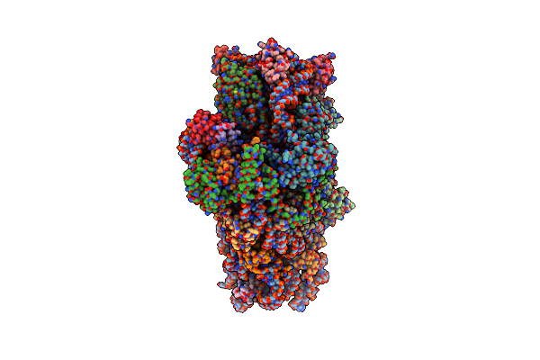
Cryo-Em Structure Of Leishmania Major 80S Ribosome With A/P/E-Site Trna And Mrna : Lm32Cs1C1 M2 Oe Mutant
Organism: Leishmania major strain friedlin
Method: ELECTRON MICROSCOPY Release Date: 2025-07-23 Classification: RIBOSOME Ligands: SPD, MG, NA, K, PUT, PAR, ZN |
 |
Method: ELECTRON MICROSCOPY
Release Date: 2025-05-28 Classification: RIBOSOME Ligands: MG, SPD, PUT, SPM, ZN |
 |
Method: ELECTRON MICROSCOPY
Release Date: 2025-05-28 Classification: RIBOSOME Ligands: MG, SPD, SPM, PUT, ZN |
 |
Staphylococcus Aureus Fusb Bound To The Small Subunit Of The S. Aureus 70S Ribosome (Fusb-Sa70S:Ssu)
Organism: Staphylococcus aureus, Escherichia coli k-12, Staphylococcus aureus subsp. aureus nctc 8325
Method: ELECTRON MICROSCOPY Resolution:2.22 Å Release Date: 2025-03-26 Classification: RIBOSOME Ligands: ZN, MG, PUT |
 |
Staphylococcus Aureus Fusb Bound To The Large Subunit Of The S. Aureus 70S Ribosome (Fusb-Sa70S:Lsu)
Organism: Staphylococcus aureus, Escherichia coli k-12, Staphylococcus aureus subsp. aureus nctc 8325
Method: ELECTRON MICROSCOPY Resolution:2.70 Å Release Date: 2025-03-26 Classification: RIBOSOME Ligands: ZN, MG, PUT |
 |
Organism: Escherichia coli
Method: ELECTRON MICROSCOPY Release Date: 2025-03-19 Classification: RIBOSOME Ligands: MG, K, SPD, PUT, ZN |
 |
Organism: Escherichia coli
Method: ELECTRON MICROSCOPY Release Date: 2025-03-19 Classification: RIBOSOME Ligands: MG, K, SPD, PUT, ZN |
 |
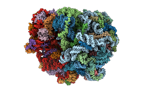
Impacts Of Ribosomal Rna Sequence Variation On Gene Expression And Phenotype: Cryo-Em Structure Of The Rrsb Ribosome (Bbb-70S)
Organism: Escherichia coli k-12, Escherichia coli bl21
Method: ELECTRON MICROSCOPY Release Date: 2025-03-19 Classification: RIBOSOME Ligands: PUT, SPD, MG, K, ZN, ATP, FME |
 |
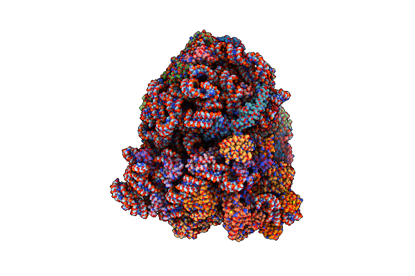
Impacts Of Ribosomal Rna Sequence Variation On Gene Expression And Phenotype: Cryo-Em Structure Of The Rrsh Ribosome (Hbb-70S)
Organism: Escherichia coli k-12, Escherichia coli bl21
Method: ELECTRON MICROSCOPY Release Date: 2025-03-19 Classification: RIBOSOME Ligands: PUT, SPD, MG, K, ZN, ATP, FME |
 |
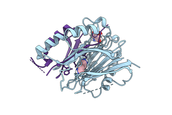
Crystal Structure Of The Engineered C-Terminal Phosphatase Domain From Saccharomyces Cerevisiae Vip1 In Complex With 1,5-Insp8 (Phosphatase Dead Mutant, Loop Deletion Residues 848-918)
Organism: Saccharomyces cerevisiae
Method: X-RAY DIFFRACTION Resolution:2.36 Å Release Date: 2025-02-26 Classification: BIOSYNTHETIC PROTEIN Ligands: ZN, SPM, ORN, PUT, EDO, I8P, 1JW |
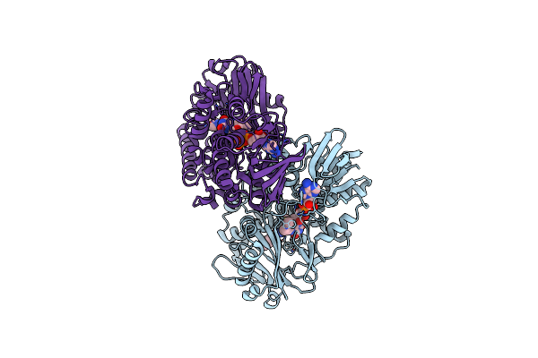 |
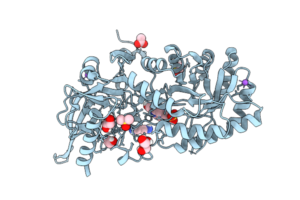
Crystal Structure Of Thermoanaerobacterales Bacterium Monoamine Oxidase In Complex With Putrescine
Organism: Thermoanaerobacterales bacterium
Method: X-RAY DIFFRACTION Resolution:2.30 Å Release Date: 2025-01-22 Classification: FLAVOPROTEIN Ligands: FAD, MG, PUT |
 |
Organism: Homo sapiens
Method: ELECTRON MICROSCOPY Release Date: 2024-12-11 Classification: RIBOSOME Ligands: MG, PUT, K, ZN, SPD, SPM, ATP |
 |
Mycoplasma Pneumoniae Small Ribosomal Subunit In Chloramphenicol-Treated Cells
Organism: Mycoplasmoides pneumoniae m129
Method: ELECTRON MICROSCOPY Release Date: 2024-11-20 Classification: TRANSLATION Ligands: SPD, PUT, N2P, MG, ZN |