
Search Count: 95
All
Selected
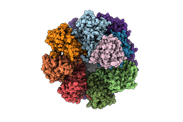 |
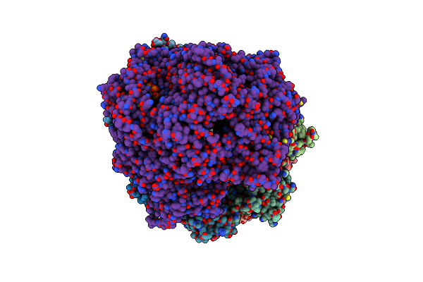
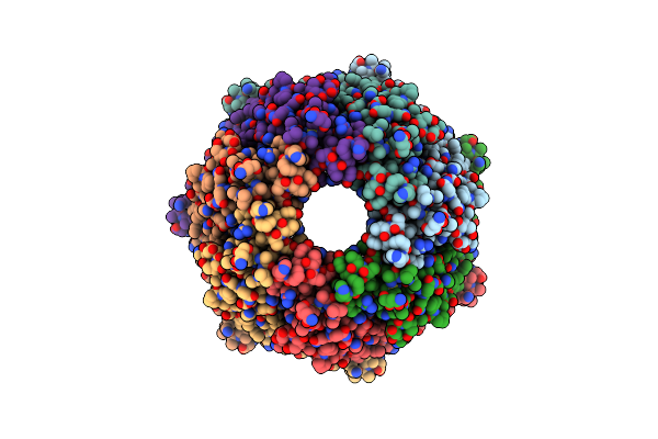
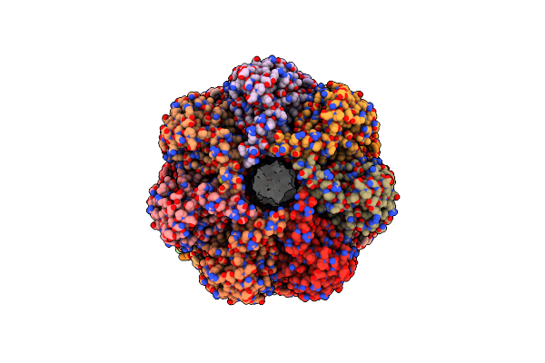
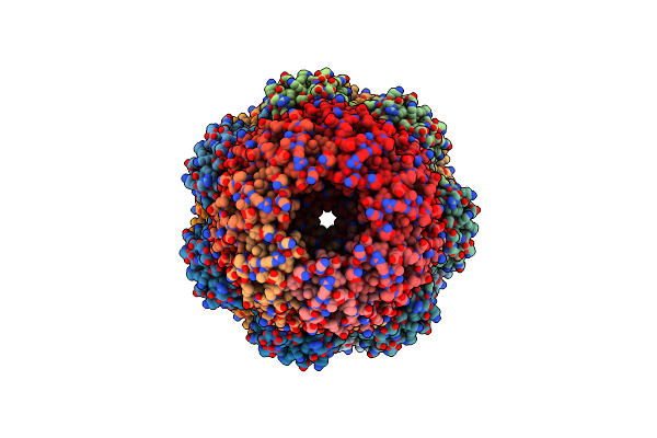
Proteasome Core Particle Assembly Intermediate Blm10:Alpha-Ring Purified From Saccharomyces Cerevisiae.
Organism: Saccharomyces cerevisiae
Method: ELECTRON MICROSCOPY Resolution:2.75 Å Release Date: 2025-08-20 Classification: HYDROLASE |
 |
Cryo-Em Structure Of The Bacterial Proteasome Activator Bpa Of Mycobacterium Tuberculosis
Organism: Mycobacterium tuberculosis h37rv
Method: ELECTRON MICROSCOPY Release Date: 2025-04-02 Classification: CHAPERONE |
 |
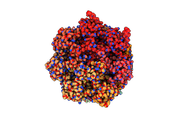
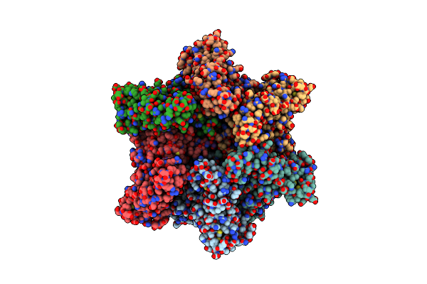
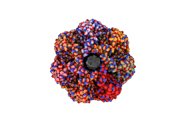
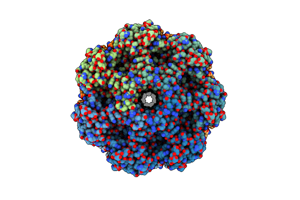
Proteasome Core Particle Assembly Intermediate Blm10:13S Purified From Saccharomyces Cerevisiae
Organism: Saccharomyces cerevisiae
Method: ELECTRON MICROSCOPY Resolution:2.84 Å Release Date: 2024-12-04 Classification: HYDROLASE |
 |
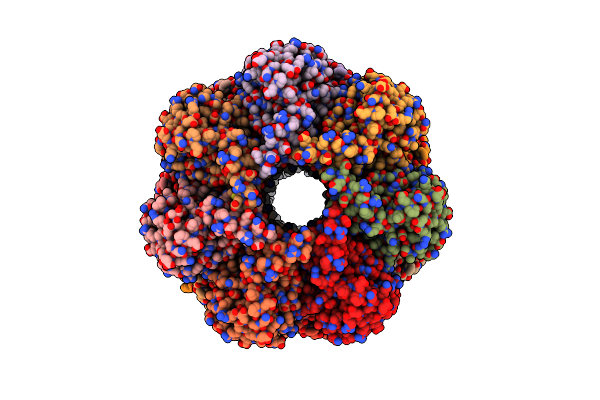
Organism: Saccharomyces cerevisiae
Method: ELECTRON MICROSCOPY Release Date: 2024-09-11 Classification: HYDROLASE |
 |
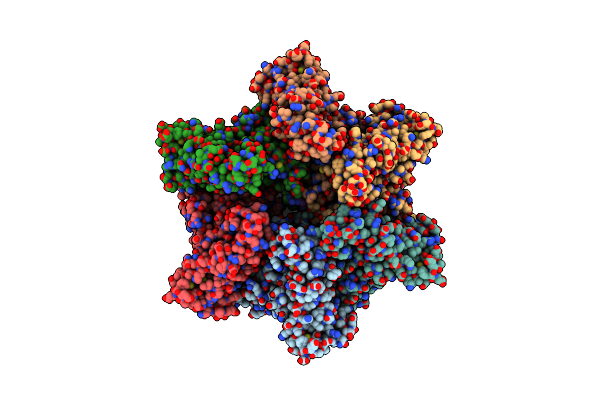
Organism: Homo sapiens
Method: ELECTRON MICROSCOPY Release Date: 2022-11-02 Classification: HYDROLASE |
 |
Organism: Homo sapiens
Method: ELECTRON MICROSCOPY Release Date: 2022-11-02 Classification: HYDROLASE |
 |
Organism: Homo sapiens
Method: ELECTRON MICROSCOPY Release Date: 2022-11-02 Classification: HYDROLASE Ligands: IHP |
 |
Organism: Homo sapiens
Method: ELECTRON MICROSCOPY Release Date: 2022-11-02 Classification: HYDROLASE/INHIBITOR Ligands: LDZ, IHP |
 |
Organism: Homo sapiens
Method: ELECTRON MICROSCOPY Release Date: 2022-11-02 Classification: HYDROLASE |
 |
Organism: Homo sapiens
Method: ELECTRON MICROSCOPY Release Date: 2022-09-21 Classification: PROTEIN TRANSPORT |
 |
Organism: Homo sapiens
Method: ELECTRON MICROSCOPY Release Date: 2022-09-21 Classification: PROTEIN TRANSPORT |
 |
Substrate-Engaged Mycobacterial Proteasome-Associated Atpase - Focused 3D Refinement (State A)
Organism: Mycobacterium tuberculosis
Method: ELECTRON MICROSCOPY Release Date: 2022-01-19 Classification: CYTOSOLIC PROTEIN Ligands: ATP, MG, ADP |
 |
Open-Gate Mycobacterium 20S Cp Proteasome In Complex Mpa - Global 3D Refinement
Organism: Mycobacterium tuberculosis
Method: ELECTRON MICROSCOPY Release Date: 2022-01-19 Classification: CYTOSOLIC PROTEIN |
 |
Substrate-Engaged Mycobacterial Proteasome-Associated Atpase - Focused 3D Refinement (State B)
Organism: Mycobacterium tuberculosis
Method: ELECTRON MICROSCOPY Release Date: 2022-01-19 Classification: CHAPERONE Ligands: ATP, MG |
 |
Substrate-Engaged Mycobacterial Proteasome-Associated Atpase In Complex With Open-Gate 20S Cp - Composite Map (State A)
Organism: Mycobacterium tuberculosis (strain atcc 25618 / h37rv)
Method: ELECTRON MICROSCOPY Release Date: 2022-01-19 Classification: CYTOSOLIC PROTEIN Ligands: ATP, MG, ADP |
 |
Substrate-Engaged Mycobacterial Proteasome-Associated Atpase In Complex With Open-Gate 20S Cp - Composite Map (State B)
Organism: Mycobacterium tuberculosis
Method: ELECTRON MICROSCOPY Release Date: 2022-01-19 Classification: CYTOSOLIC PROTEIN Ligands: ATP, MG |
 |
Organism: Homo sapiens, Bos taurus
Method: ELECTRON MICROSCOPY Release Date: 2021-01-20 Classification: HYDROLASE |
 |
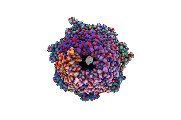
Bovine 20S Immunoproteasome In Complex With Two Human Pa28Alpha-Beta Activators
Organism: Homo sapiens, Bos taurus
Method: ELECTRON MICROSCOPY Release Date: 2021-01-20 Classification: HYDROLASE |
 |
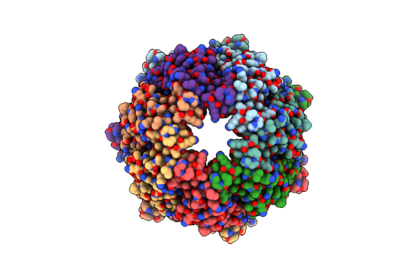
Allosteric Coupling Between Alpha-Rings Of The 20S Proteasome, 20S Singly Capped With A Pa26/V230F
Organism: Thermoplasma acidophilum, Trypanosoma brucei
Method: ELECTRON MICROSCOPY Release Date: 2020-09-09 Classification: HYDROLASE |
 |
Allosteric Coupling Between Alpha-Rings Of 20S Proteasome, 20S Proteasome Singly Capped With A Pa26/E102A_Panc, Together With Lfp Incubation
Organism: Thermoplasma acidophilum, Trypanosoma brucei brucei
Method: ELECTRON MICROSCOPY Release Date: 2020-09-09 Classification: HYDROLASE |