
Search Count: 207
 |
Organism: Homo sapiens, Synthetic construct
Method: X-RAY DIFFRACTION Release Date: 2025-12-31 Classification: TRANSFERASE Ligands: CL, PRO, PEG |
 |
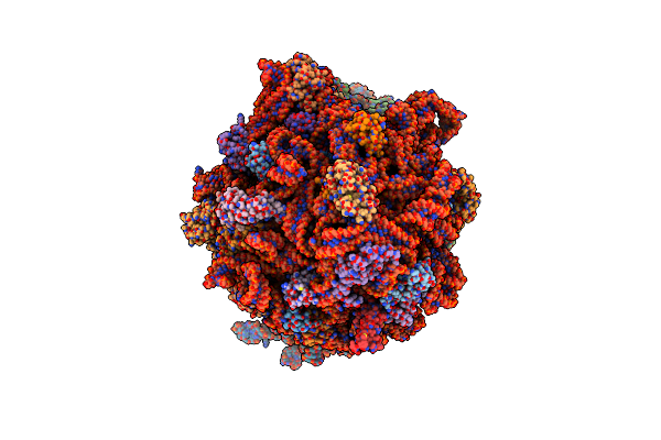
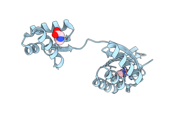
Variant T321N Of Orotidine 5'-Monophosphate Decarboxylase-Domain Of Human Umps In Complex With The Product Ump And The Nucleotide Tmp At 0.86 Angstrom Resolution
Organism: Homo sapiens
Method: X-RAY DIFFRACTION Release Date: 2025-10-22 Classification: LYASE Ligands: U5P, PRO, TMP |
 |
Variant T321N Of Orotidine 5'-Monophosphate Decarboxylase-Domain Of Human Umps In Complex With Deoxyuridine-5'-Monophosphate (Deoxy Ump) At 1.3 Angstroms Resolution
Organism: Homo sapiens
Method: X-RAY DIFFRACTION Release Date: 2025-10-22 Classification: LYASE Ligands: DU, PRO |
 |
Orotidine 5'-Monophosphate Decarboxylase-Domain Of Human Umps In Complex With Xmp At 1.0 Angstrom Resolution
Organism: Homo sapiens
Method: X-RAY DIFFRACTION Release Date: 2025-10-22 Classification: LYASE Ligands: XMP, PRO |
 |
Orotidine 5'-Monophosphate Decarboxylase-Domain Of Human Umps In Complex With A New Inhibitor 1-(Beta-D-Ribofuranosyl) Cyanuric Acid-5'-Monophosphate (Ymp)
Organism: Homo sapiens
Method: X-RAY DIFFRACTION Release Date: 2025-10-22 Classification: LYASE Ligands: A1IT4, PRO |
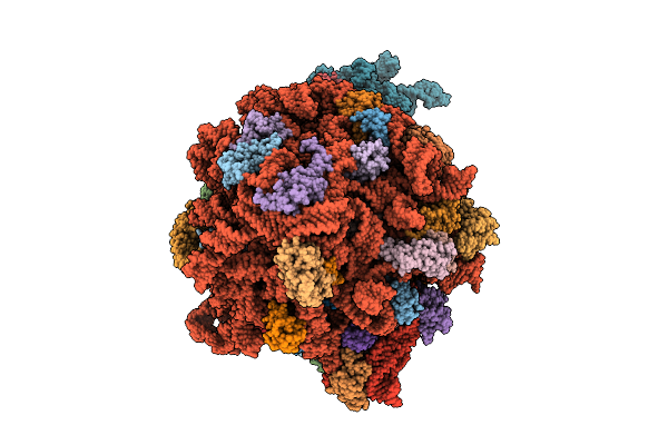 |
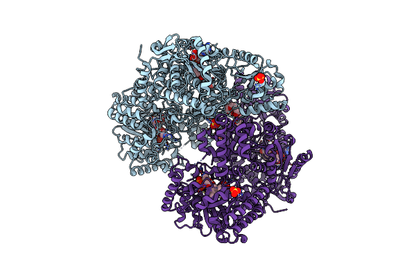
Organism: Escherichia coli
Method: ELECTRON MICROSCOPY Release Date: 2025-10-22 Classification: RIBOSOME Ligands: ZN, PRO |
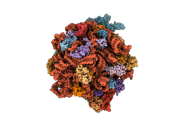 |
Structure Of E.Coli Ribosome In Complex With An Engineered Arrest Peptide And Trigger Factor
Organism: Escherichia coli
Method: ELECTRON MICROSCOPY Release Date: 2025-10-22 Classification: RIBOSOME Ligands: ZN, PRO |
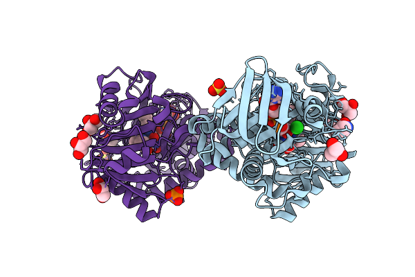 |
Monomeric Sarcosine Oxidase From Bacillus Sp. (Soxb) Complexed With L-Proline
Organism: Bacillus sp. b-0618
Method: X-RAY DIFFRACTION Release Date: 2025-09-24 Classification: OXIDOREDUCTASE Ligands: FAD, PRO, GOL, CL, SO4, PO4 |
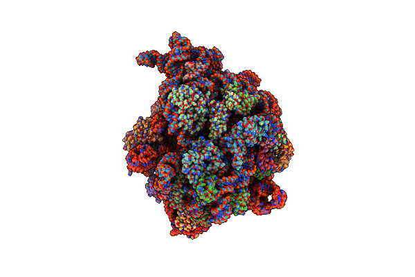 |
Organism: Escherichia coli
Method: ELECTRON MICROSCOPY Release Date: 2025-06-18 Classification: RIBOSOME Ligands: ZN, PRO |
 |
Structure Of E.Coli Ribosome In Complex With An Engineered Arrest Peptide And Trigger Factor
Organism: Escherichia coli
Method: ELECTRON MICROSCOPY Release Date: 2025-06-18 Classification: RIBOSOME Ligands: ZN, PRO |
 |
Organism: Escherichia coli (strain k12)
Method: ELECTRON MICROSCOPY Release Date: 2025-06-04 Classification: TRANSFERASE Ligands: PRO |
 |
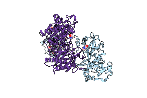
Proline Utilization A With The Fadh- N5 Atom Covalently Modified By Proline
Organism: Sinorhizobium meliloti sm11
Method: X-RAY DIFFRACTION Release Date: 2025-06-04 Classification: OXIDOREDUCTASE Ligands: NAD, PGE, FDA, A1CAB, PRO, MG, SO4, A1AT4 |
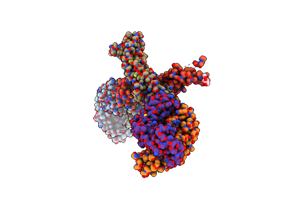 |
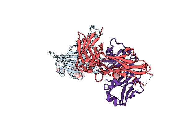
Fertilization Izumo1 Protein Ectodomain In Complex With Anti-Sperm Antibody Obf13
Organism: Mus musculus
Method: X-RAY DIFFRACTION Resolution:3.18 Å Release Date: 2025-03-19 Classification: CELL ADHESION Ligands: NAG, PRO, CL |
 |
Organism: Homo sapiens
Method: X-RAY DIFFRACTION Resolution:1.44 Å Release Date: 2025-01-22 Classification: IMMUNE SYSTEM Ligands: TB, 9JE, BTB, PRO, YT3, PG4, ER3 |
 |
Organism: Thermotoga maritima
Method: X-RAY DIFFRACTION Resolution:2.30 Å Release Date: 2025-01-22 Classification: DNA BINDING PROTEIN Ligands: FE2, NIO, PRO |
 |
Organism: Paracoccus denitrificans pd1222
Method: X-RAY DIFFRACTION Resolution:1.45 Å Release Date: 2025-01-01 Classification: LYASE Ligands: ZN, PRO |
 |
Organism: Homo sapiens
Method: X-RAY DIFFRACTION Resolution:1.90 Å Release Date: 2024-12-04 Classification: HYDROLASE Ligands: MN, DMS, PRO |
 |
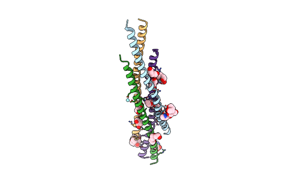
Organism: Pelagibacter sp. (strain htcc7211)
Method: X-RAY DIFFRACTION Resolution:1.30 Å Release Date: 2024-07-03 Classification: TRANSPORT PROTEIN Ligands: PRO, MES, PG4, GOL |
 |
Organism: Severe acute respiratory syndrome coronavirus 2, Homo sapiens
Method: X-RAY DIFFRACTION Release Date: 2024-06-19 Classification: VIRAL PROTEIN Ligands: PRO, GOL |
 |
Crystal Structure Of Apo Form Of Short Prokaryotic Argonaute Tir-Apaz (Sparta) Heterodimer
Organism: Thermoflavifilum thermophilum
Method: X-RAY DIFFRACTION Resolution:2.66 Å Release Date: 2024-06-12 Classification: RNA BINDING PROTEIN Ligands: PRO, MN |