
Search Count: 58
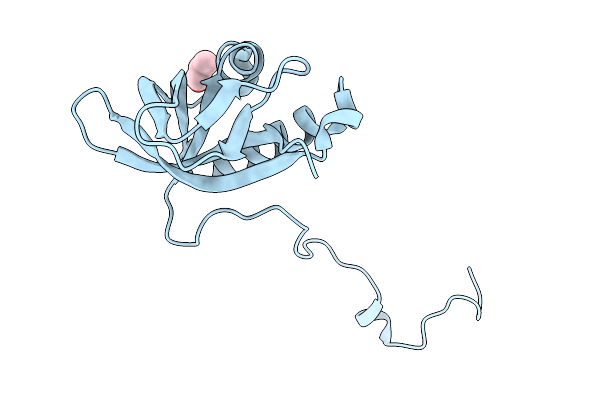 |
Organism: Paulinella chromatophora
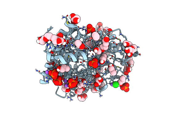
Method: X-RAY DIFFRACTION Release Date: 2025-11-19 Classification: UNKNOWN FUNCTION Ligands: POL |
 |
Crystal Structure Of Bf3526 Peptidase From Bacteroides Fragilis In Complex With A Peptide
Organism: Bacteroides fragilis nctc 9343, Synthetic construct
Method: X-RAY DIFFRACTION Release Date: 2025-07-16 Classification: HYDROLASE Ligands: K, ZN, EDO, POL, PEG, BU1, HEZ |
 |
Computationally Designed Tunable C2 Symmetric Tandem Repeat Homodimer, D_3_633_8X Bound To Peptide
Organism: Synthetic construct
Method: X-RAY DIFFRACTION Resolution:2.05 Å Release Date: 2024-12-11 Classification: DE NOVO PROTEIN Ligands: POL, SO4 |
 |
The Crystal Structure Of Protein A21, A Component Of The Conserved Poxvirus Entry-Fusion Complex
Organism: Vaccinia virus western reserve
Method: X-RAY DIFFRACTION Resolution:2.30 Å Release Date: 2024-09-04 Classification: VIRAL PROTEIN Ligands: EOH, EDO, MOH, IOD, CL, PDO, PGR, NA, PG0, DHL, PGE, PEG, POL, GOL, IPA |
 |
Organism: Thomasclavelia ramosa
Method: X-RAY DIFFRACTION Resolution:1.70 Å Release Date: 2024-07-24 Classification: ANTITOXIN Ligands: POL, CAC, GOL, MG, APC |
 |
Organism: Homo sapiens
Method: X-RAY DIFFRACTION Resolution:2.50 Å Release Date: 2023-06-14 Classification: PROTEIN TRANSPORT Ligands: POL |
 |
Organism: Synthetic construct
Method: X-RAY DIFFRACTION Resolution:1.68 Å Release Date: 2022-07-20 Classification: DE NOVO PROTEIN Ligands: POL, SO4 |
 |
Organism: Clostridioides difficile (strain r20291)
Method: X-RAY DIFFRACTION Resolution:2.20 Å Release Date: 2022-03-23 Classification: PEPTIDE BINDING PROTEIN Ligands: SO4, EPE, POL, PEG, ZN, ZZ7 |
 |
Sars-Cov-2 Spike Receptor-Binding Domain (Rbd) In Complex With S2X35 Fab And S309 Fab
Organism: Homo sapiens, Severe acute respiratory syndrome coronavirus 2
Method: X-RAY DIFFRACTION Resolution:1.83 Å Release Date: 2021-07-21 Classification: VIRAL PROTEIN/IMMUNE SYSTEM Ligands: SO4, GOL, CL, POL |
 |
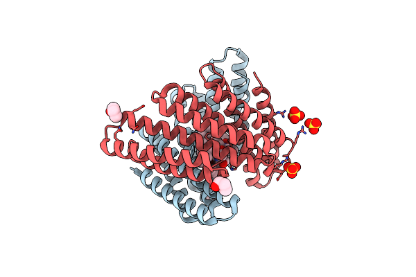
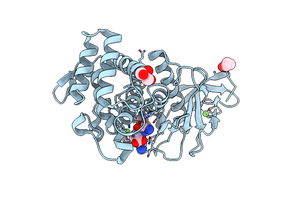
Nitric Oxide Synthase From Bacillus Subtilis In Complex With 7-((3-(2-(6-Aminopyridin-2-Yl)Ethyl)Phenoxy)Methyl)Quinolin-2-Amine
Organism: Bacillus subtilis (strain 168)
Method: X-RAY DIFFRACTION Resolution:1.85 Å Release Date: 2021-07-07 Classification: OXIDOREDUCTASE/Inhibitor Ligands: HEM, V5G, CL, GOL, POL |
 |
Nitric Oxide Synthase From Bacillus Subtilis In Complex With 7-((3-(((Pyridin-2-Ylmethyl)Amino)Methyl)Phenoxy)Methyl)Quinolin-2-Amine
Organism: Bacillus subtilis (strain 168)
Method: X-RAY DIFFRACTION Resolution:1.95 Å Release Date: 2021-06-30 Classification: OXIDOREDUCTASE/Inhibitor Ligands: HEM, POL, GOL, V4V, CL, PEG |
 |
Nitric Oxide Synthase From Bacillus Subtilis In Complex With 7-((3-(2-(6-Aminopyridin-2-Yl)Ethyl)Phenoxy)Methyl)Quinolin-2-Amine
Organism: Bacillus subtilis (strain 168)
Method: X-RAY DIFFRACTION Resolution:1.85 Å Release Date: 2021-06-30 Classification: OXIDOREDUCTASE/Inhibitor Ligands: HEM, V5G, CL, GOL, POL |
 |
Structure Of Bacillus Subtilis Nitric Oxide Synthase In Complex With 7-((3-((((6-Aminopyridin-2-Yl)Methyl)Amino)Methyl)Phenoxy)Methyl)Quinolin-2-Amine
Organism: Bacillus subtilis (strain 168)
Method: X-RAY DIFFRACTION Resolution:2.25 Å Release Date: 2021-06-30 Classification: OXIDOREDUCTASE/Inhibitor Ligands: HEM, V54, CL, GOL, PEG, POL |
 |
Nitric Oxide Synthase From Bacillus Subtilis In Complex With N2-((3-((2-Aminoquinolin-7-Yl)Methoxy)Phenoxy)Methyl)Pyridine-2,6-Diamine
Organism: Bacillus subtilis (strain 168)
Method: X-RAY DIFFRACTION Resolution:2.25 Å Release Date: 2021-06-16 Classification: OXIDOREDUCTASE Ligands: HEC, CL, GOL, POL, PEG, V0P |
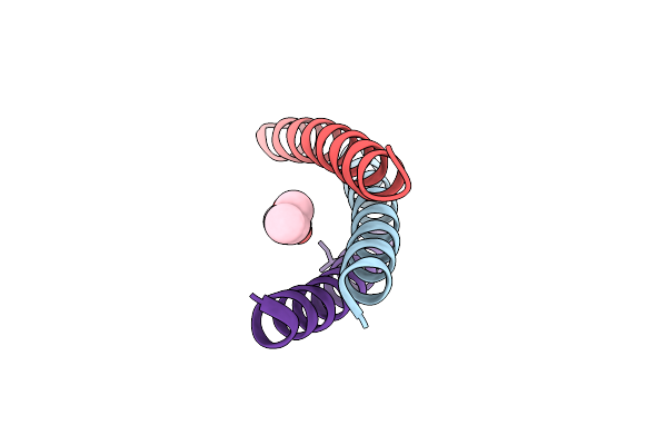 |
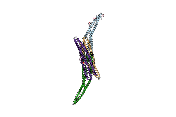
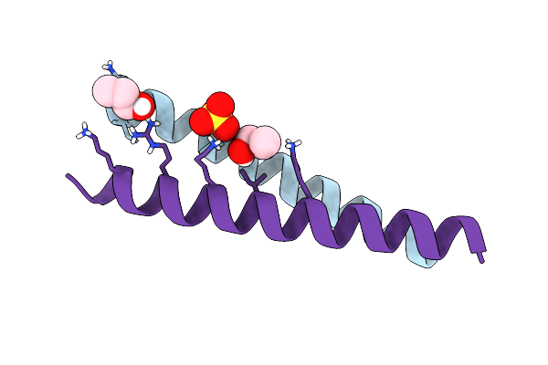
A Hexameric Barrel State Of A De Novo Coiled-Coil Assembly: Cc-Type2-(Ggiaid)4
Organism: Synthetic construct
Method: X-RAY DIFFRACTION Resolution:1.77 Å Release Date: 2021-04-28 Classification: DE NOVO PROTEIN Ligands: POL |
 |
A Heptameric Barrel State Of A De Novo Coiled-Coil Assembly: Cc-Type2-(Ggiaid)4
Organism: Synthetic construct
Method: X-RAY DIFFRACTION Resolution:1.71 Å Release Date: 2021-04-28 Classification: DE NOVO PROTEIN Ligands: POL, GOL |
 |
Crystal Structure Of The Deah-Box Atpase Prp2 In Complex With Adp-Bef3 And Ssrna
Organism: Chaetomium thermophilum (strain dsm 1495 / cbs 144.50 / imi 039719), Synthetic construct
Method: X-RAY DIFFRACTION Resolution:2.10 Å Release Date: 2021-04-21 Classification: HYDROLASE Ligands: BEF, ADP, MG, PEG, GOL, MPO, CL, PGO, HEZ, POL, EDO |
 |
Organism: Bacillus thermoproteolyticus
Method: X-RAY DIFFRACTION Resolution:1.50 Å Release Date: 2021-01-27 Classification: HYDROLASE Ligands: ZN, CA, POL, GOL, ILE, LYS |
 |
Organism: Homo sapiens
Method: X-RAY DIFFRACTION Resolution:1.81 Å Release Date: 2020-02-05 Classification: HYDROLASE Ligands: GOL, CL, NA, PO4, POL, PGE, EOH, EDO |
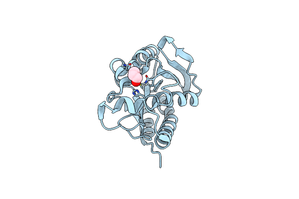 |
Crystal Structure Of The Complex Of Peptidyl-Trna Hydrolase With N-Propanol At 1.43 A Resolution
Organism: Acinetobacter baumannii (strain atcc 19606 / dsm 30007 / cip 70.34 / jcm 6841 / nbrc 109757 / ncimb 12457 / nctc 12156 / 81)
Method: X-RAY DIFFRACTION Resolution:1.43 Å Release Date: 2018-12-26 Classification: HYDROLASE Ligands: POL |