
Search Count: 106
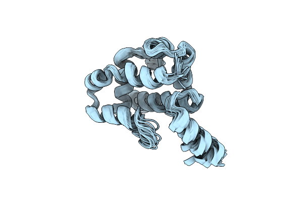 |
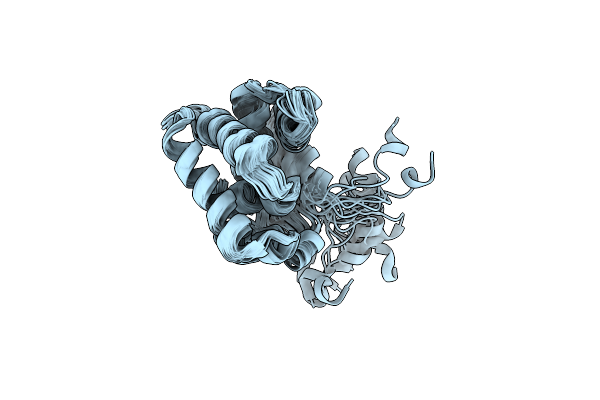
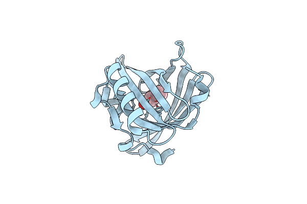
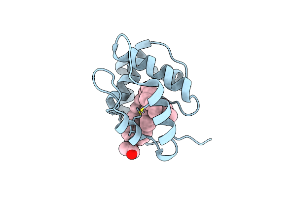
Organism: Ostrinia furnacalis
Method: SOLUTION NMR Release Date: 2023-02-15 Classification: SIGNALING PROTEIN |
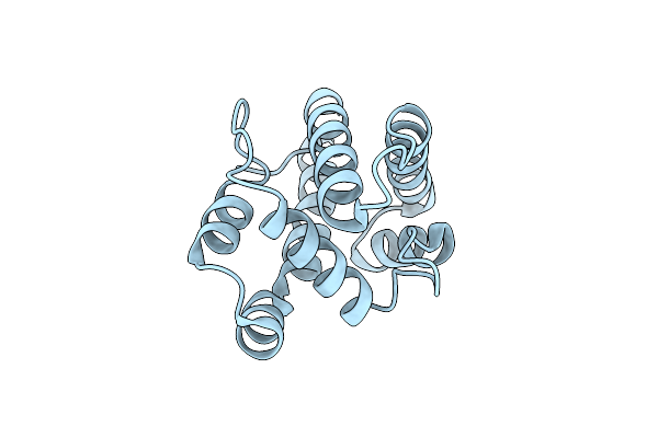 |
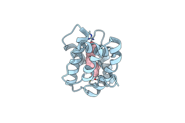
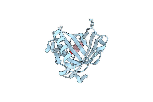
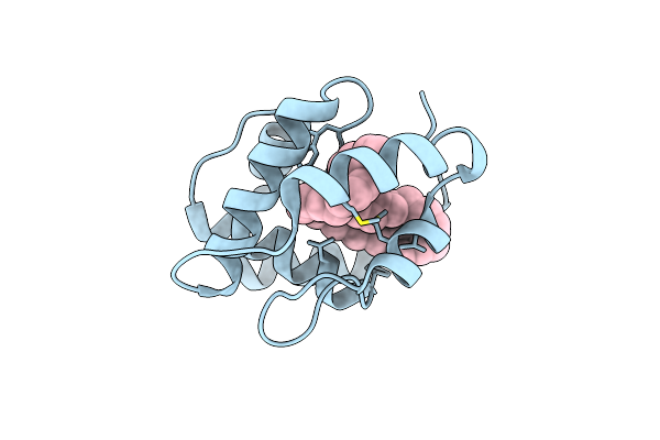
Organism: Helicoverpa armigera
Method: X-RAY DIFFRACTION Resolution:1.30 Å Release Date: 2022-02-02 Classification: TRANSPORT PROTEIN |
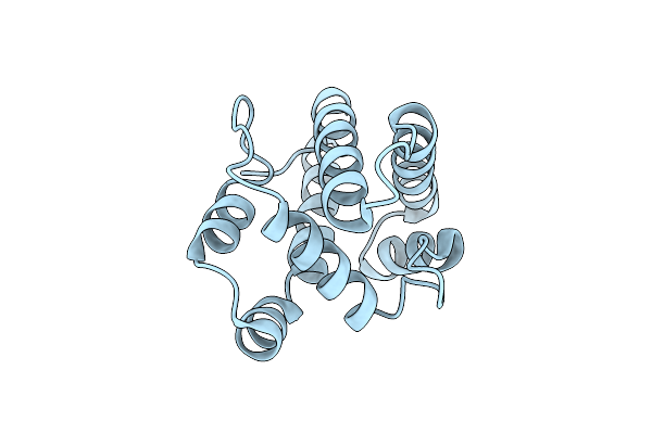 |
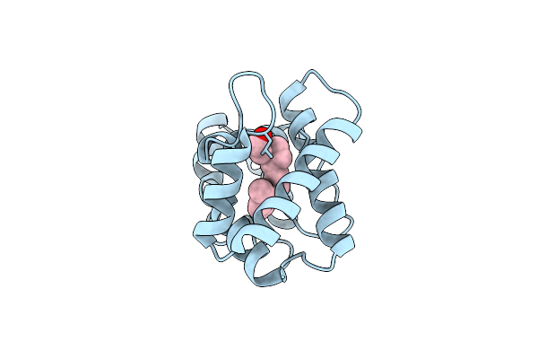
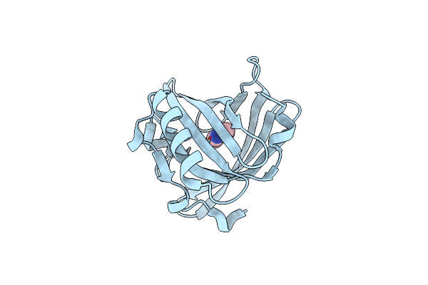
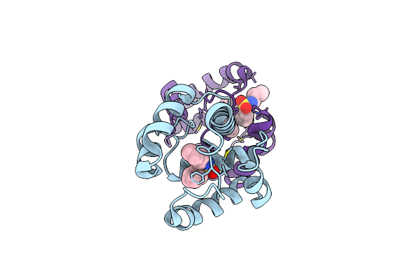
Organism: Helicoverpa armigera
Method: X-RAY DIFFRACTION Resolution:2.05 Å Release Date: 2022-02-02 Classification: TRANSPORT PROTEIN |
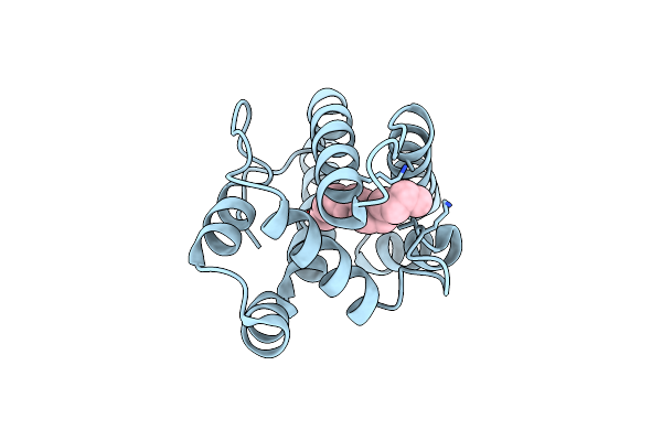 |
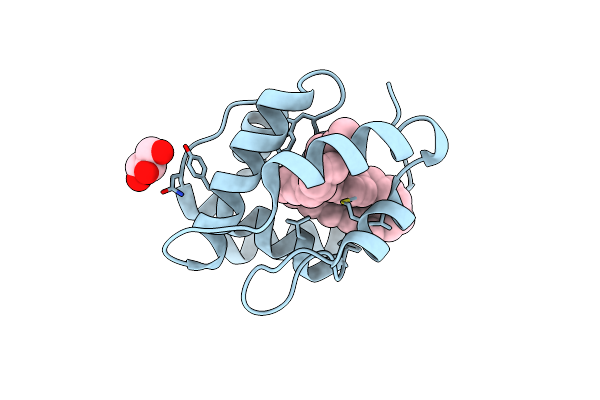
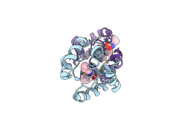
Organism: Helicoverpa armigera
Method: X-RAY DIFFRACTION Resolution:2.10 Å Release Date: 2022-02-02 Classification: TRANSPORT PROTEIN Ligands: 81K |
 |
Organism: Lymantria dispar
Method: SOLUTION NMR Release Date: 2020-09-23 Classification: LIPID BINDING PROTEIN |
 |
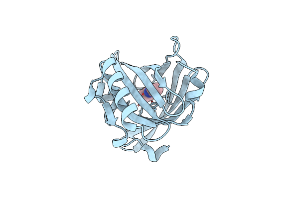
Organism: Drosophila melanogaster
Method: X-RAY DIFFRACTION Resolution:2.00 Å Release Date: 2020-03-18 Classification: TRANSPORT PROTEIN Ligands: 1PE, MLA |
 |
Structure Of Pheromone-Binding Protein 1 In Complex With (11Z,13Z)-Hexadecadienal
Organism: Amyelois transitella
Method: X-RAY DIFFRACTION Resolution:1.14 Å Release Date: 2013-03-06 Classification: pheromone-binding protein Ligands: 1EY |
 |
Structure Of Pheromone-Binding Protein 1 In Complex With (Z,Z)-11,13- Hexadecadienol
Organism: Amyelois transitella
Method: X-RAY DIFFRACTION Resolution:1.85 Å Release Date: 2013-03-06 Classification: pheromone-binding protein Ligands: 1EX |
 |
Major Mouse Urinary Protein Iv Complexed With 2-Sec-Butyl-4,5-Dihydrothiazole
Organism: Mus musculus
Method: X-RAY DIFFRACTION Resolution:0.96 Å Release Date: 2010-06-16 Classification: TRANSPORT PROTEIN Ligands: CL, XBT, ZBT |
 |
Organism: Mus musculus
Method: X-RAY DIFFRACTION Resolution:1.43 Å Release Date: 2010-06-16 Classification: TRANSPORT PROTEIN Ligands: CL, 2EH, HTX |
 |
Organism: Mus musculus
Method: X-RAY DIFFRACTION Resolution:1.02 Å Release Date: 2010-06-16 Classification: TRANSPORT PROTEIN Ligands: CL, 2EH |
 |
Organism: Mus musculus
Method: X-RAY DIFFRACTION Resolution:1.42 Å Release Date: 2010-06-16 Classification: TRANSPORT PROTEIN Ligands: CL, 25R |
 |
Crystal Structure Of A Pheromone Binding Protein From Apis Mellifera With A Serendipitous Ligand At Ph 5.5
Organism: Apis mellifera
Method: X-RAY DIFFRACTION Resolution:1.80 Å Release Date: 2009-12-01 Classification: Pheromone binding protein Ligands: CMJ, GOL, CL |
 |
Crystal Structure Of A Pheromone Binding Protein From Apis Mellifera With A Serendipitous Ligand Soaked At Ph 4.0
Organism: Apis mellifera
Method: X-RAY DIFFRACTION Resolution:1.90 Å Release Date: 2009-12-01 Classification: Pheromone binding protein Ligands: CMJ, GOL, CL |
 |
Crystal Structure Of A Pheromone Binding Protein From Apis Mellifera With A Serendipitous Ligand Soaked At Ph 7.0
Organism: Apis mellifera
Method: X-RAY DIFFRACTION Resolution:1.75 Å Release Date: 2009-12-01 Classification: Pheromone binding protein Ligands: CMJ, CL |
 |
Crystal Structure Of A Pheromone Binding Protein Mutant D35A, From Apis Mellifera, At Ph 7.0
Organism: Apis mellifera
Method: X-RAY DIFFRACTION Resolution:2.03 Å Release Date: 2009-05-26 Classification: Pheromone Binding Protein Ligands: NBB |
 |
Crystal Structure Of A Pheromone Binding Protein Mutant D35A, From Apis Mellifera, Soaked At Ph 5.5
Organism: Apis mellifera
Method: X-RAY DIFFRACTION Resolution:2.10 Å Release Date: 2009-05-26 Classification: Pheromone Binding Protein Ligands: NBB |
 |
Crystal Structure Of A Pheromone Binding Protein Mutant D35N, From Apis Mellifera, At Ph 5.5
Organism: Apis mellifera
Method: X-RAY DIFFRACTION Resolution:2.30 Å Release Date: 2009-05-26 Classification: Pheromone Binding Protein Ligands: NBB |
 |
Crystal Structure Of A Pheromone Binding Protein Mutant D35N, From Apis Mellifera, Soaked At Ph 7.0
Organism: Apis mellifera
Method: X-RAY DIFFRACTION Resolution:1.90 Å Release Date: 2009-05-26 Classification: Pheromone binding protein |
 |
Crystal Structure Of A Pheromone Binding Protein Mutant D35N, From Apis Mellifera, Soaked At Ph 4.0
Organism: Apis mellifera
Method: X-RAY DIFFRACTION Resolution:1.70 Å Release Date: 2009-05-26 Classification: Pheromone binding protein |