Search Count: 222
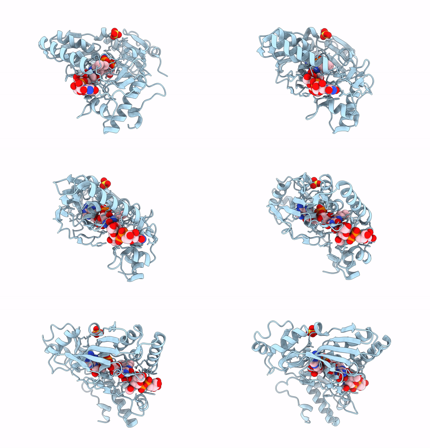 |
Crystal Structure Of Udp-N-Acetylenolpyruvoylglucosamine Reductase (Murb) From Brucella Ovis
Organism: Brucella ovis atcc 25840
Method: X-RAY DIFFRACTION Resolution:2.27 Å Release Date: 2025-01-22 Classification: OXIDOREDUCTASE Ligands: SO4, FAD, EPU |
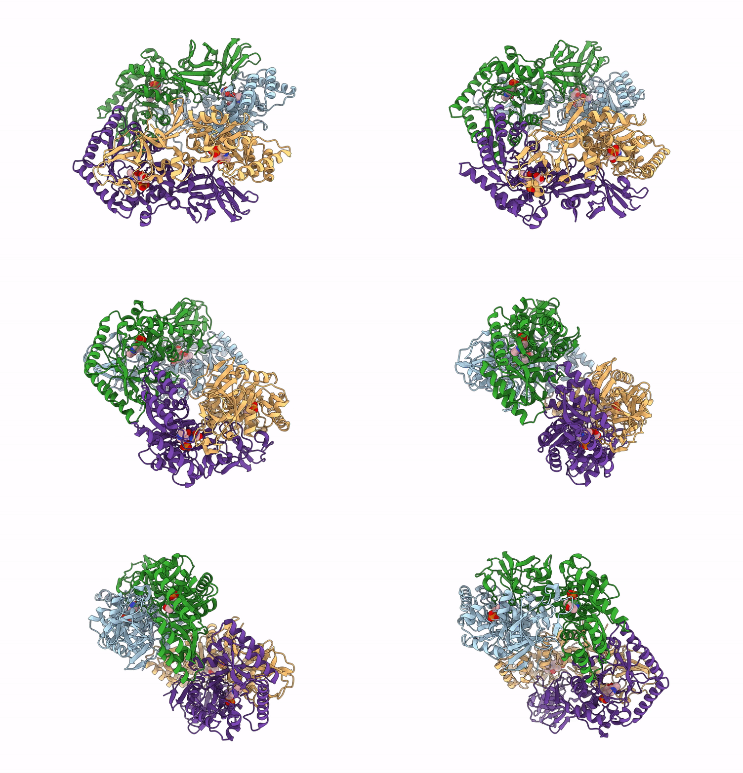 |
Organism: Bacillus subtilis subsp. subtilis str. 168
Method: X-RAY DIFFRACTION Resolution:2.30 Å Release Date: 2025-01-01 Classification: ISOMERASE Ligands: PLP, CL, ALA |
 |
Organism: Bacillus subtilis subsp. subtilis str. 168
Method: X-RAY DIFFRACTION Resolution:2.30 Å Release Date: 2025-01-01 Classification: ISOMERASE Ligands: PLP, CL, ALA |
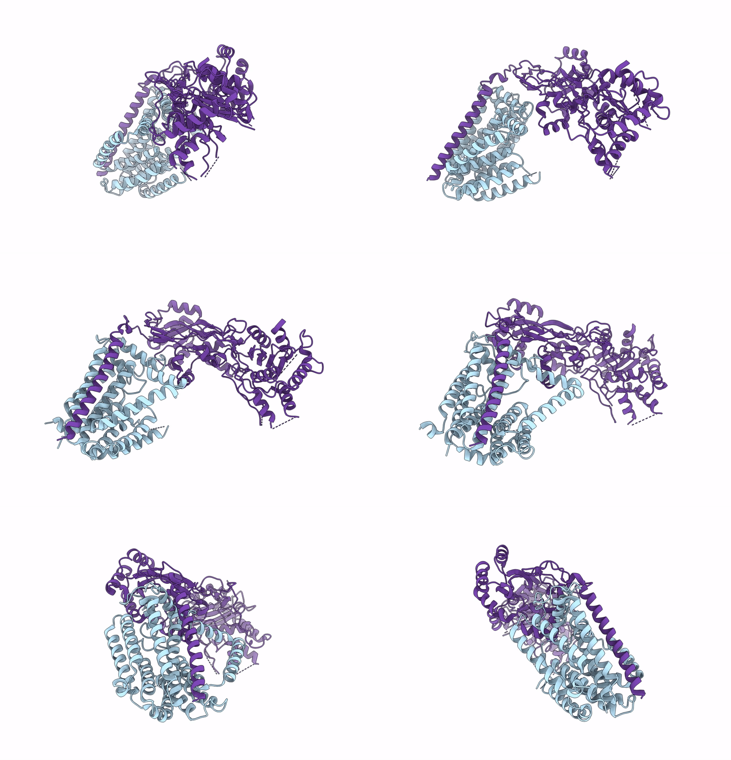 |
Organism: Escherichia coli
Method: ELECTRON MICROSCOPY Release Date: 2023-08-30 Classification: MEMBRANE PROTEIN |
 |
Organism: Pseudomonas aeruginosa pao1
Method: ELECTRON MICROSCOPY Release Date: 2023-04-19 Classification: MEMBRANE PROTEIN |
 |
Organism: Streptococcus pneumoniae r6
Method: X-RAY DIFFRACTION Resolution:1.47 Å Release Date: 2022-11-02 Classification: TRANSFERASE Ligands: MG, CL |
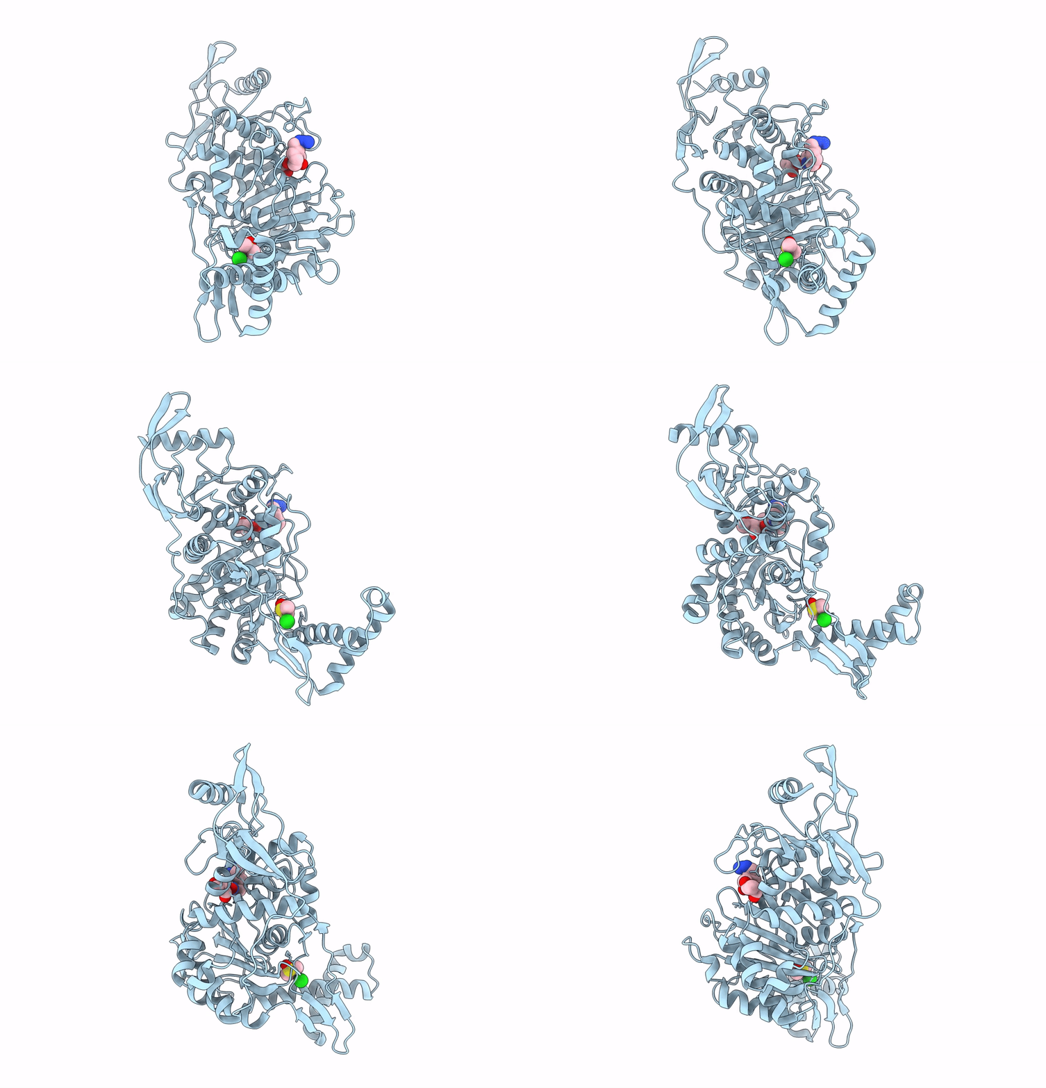 |
Penicillin-Binding Protein 1B (Pbp-1B) In Complex With Lactone 5Az - Streptococcus Pneumoniae R6
Organism: Streptococcus pneumoniae r6
Method: X-RAY DIFFRACTION Resolution:1.57 Å Release Date: 2022-11-02 Classification: TRANSFERASE Ligands: K2O, CL, DMS |
 |
Penicillin-Binding Protein 1B (Pbp-1B) In Complex With Lactone 6Az - Streptococcus Pneumoniae R6
Organism: Streptococcus pneumoniae r6
Method: X-RAY DIFFRACTION Resolution:1.55 Å Release Date: 2022-11-02 Classification: TRANSFERASE Ligands: KQN, CL, DMS |
 |
Penicillin-Binding Protein 1B (Pbp-1B) In Complex With Lactone 7Az - Streptococcus Pneumoniae R6
Organism: Streptococcus pneumoniae r6
Method: X-RAY DIFFRACTION Resolution:1.63 Å Release Date: 2022-11-02 Classification: TRANSFERASE Ligands: KQI, CL, DMS |
 |
Penicillin-Binding Protein 1B (Pbp-1B) In Complex With 8Az Lactone - Streptococcus Pneumoniae R6
Organism: Streptococcus pneumoniae r6
Method: X-RAY DIFFRACTION Resolution:1.74 Å Release Date: 2022-11-02 Classification: TRANSFERASE Ligands: JWL, CL |
 |
Crystal Structure Of Penicillin-Binding Protein 1 (Pbp1) From Staphylococcus Aureus
Organism: Staphylococcus aureus subsp. aureus col
Method: X-RAY DIFFRACTION Resolution:3.03 Å Release Date: 2021-11-03 Classification: HYDROLASE Ligands: EPE, CD, CL |
 |
Crystal Structure Of Penicillin-Binding Protein 1 (Pbp1) From Staphylococcus Aureus In Complex With Piperacillin
Organism: Staphylococcus aureus subsp. aureus col
Method: X-RAY DIFFRACTION Resolution:3.03 Å Release Date: 2021-11-03 Classification: HYDROLASE Ligands: YPP |
 |
Crystal Structure Of Penicillin-Binding Protein 1 (Pbp1) From Staphylococcus Aureus In Complex With Penicillin G
Organism: Staphylococcus aureus subsp. aureus col
Method: X-RAY DIFFRACTION Resolution:2.59 Å Release Date: 2021-11-03 Classification: HYDROLASE Ligands: CIT, SO4, PNM |
 |
Crystal Structure Of Pasta Domains Of The Penicillin-Binding Protein 1 (Pbp1) From Staphylococcus Aureus
Organism: Staphylococcus aureus subsp. aureus col
Method: X-RAY DIFFRACTION Resolution:1.51 Å Release Date: 2021-11-03 Classification: HYDROLASE Ligands: CL |
 |
Crystal Structure Of Penicillin-Binding Protein 1 (Pbp1) From Staphylococcus Aureus In Complex With Pentaglycine
Organism: Staphylococcus aureus (strain col), Synthetic construct
Method: X-RAY DIFFRACTION Resolution:3.36 Å Release Date: 2021-11-03 Classification: HYDROLASE Ligands: CD, CL |
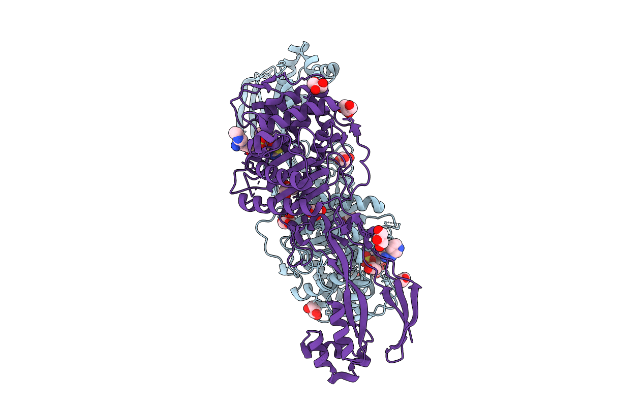 |
Organism: Pseudomonas aeruginosa (strain atcc 15692 / dsm 22644 / cip 104116 / jcm 14847 / lmg 12228 / 1c / prs 101 / pao1)
Method: X-RAY DIFFRACTION Resolution:1.73 Å Release Date: 2021-08-04 Classification: PROTEIN BINDING Ligands: VL5, CL, GOL |
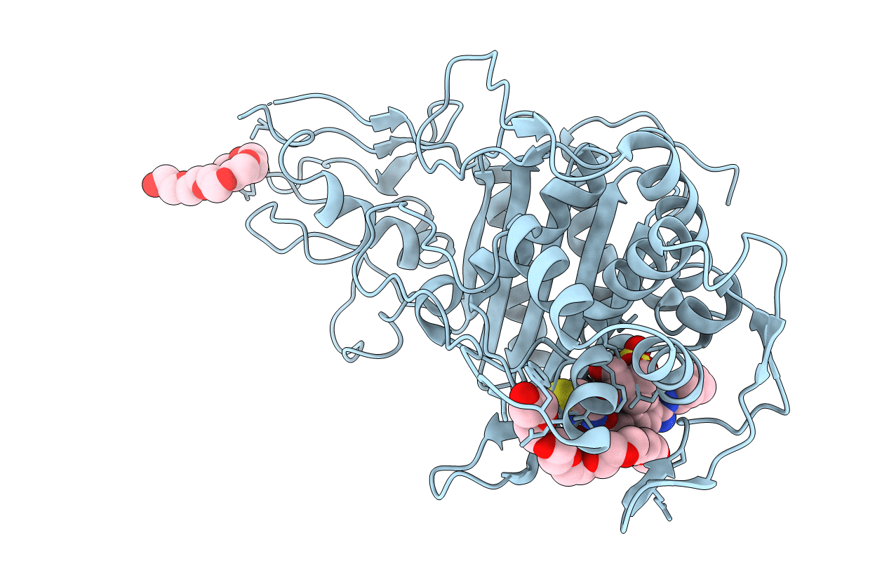 |
Crystal Structure Of Pbp3 Transpeptidase Domain From E. Coli In Complex With Aic499
Organism: Escherichia coli (strain k12)
Method: X-RAY DIFFRACTION Resolution:1.92 Å Release Date: 2021-08-04 Classification: PROTEIN BINDING Ligands: VL5, 12P |
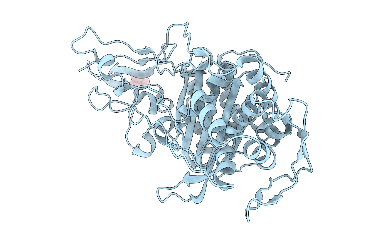 |
Organism: Escherichia coli (strain k12)
Method: X-RAY DIFFRACTION Resolution:2.30 Å Release Date: 2021-08-04 Classification: MEMBRANE PROTEIN Ligands: TMO |
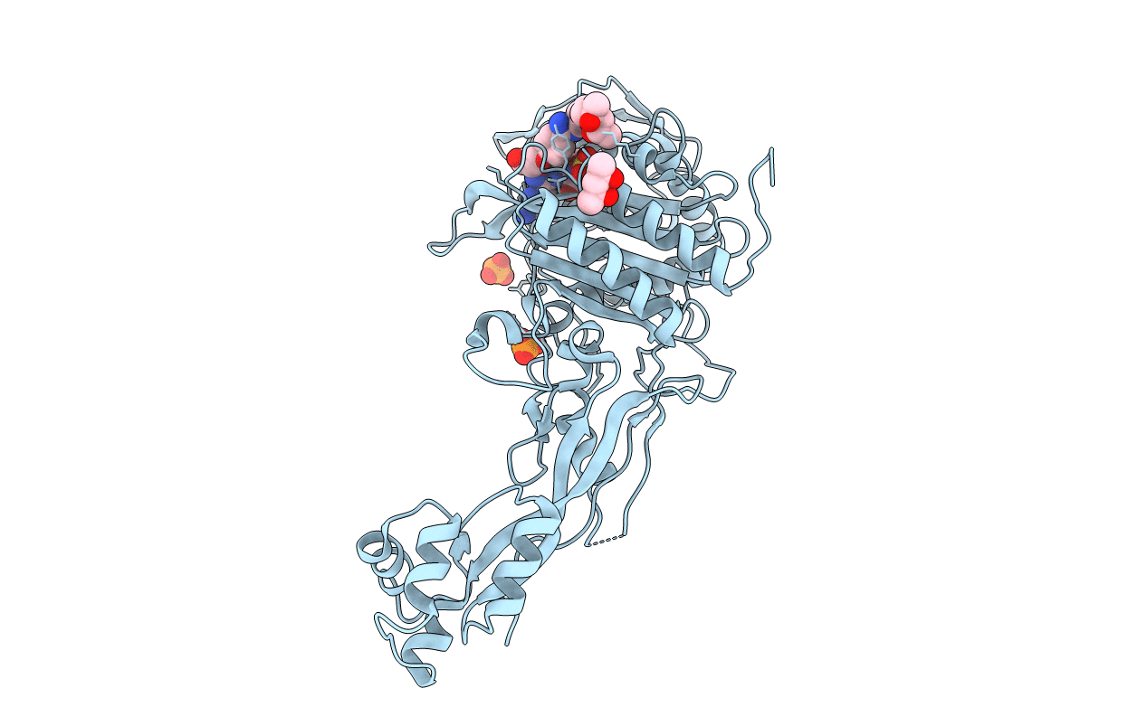 |
Organism: Escherichia coli (strain k12)
Method: X-RAY DIFFRACTION Resolution:2.70 Å Release Date: 2021-08-04 Classification: PROTEIN BINDING Ligands: VL5, PO4, MPD |
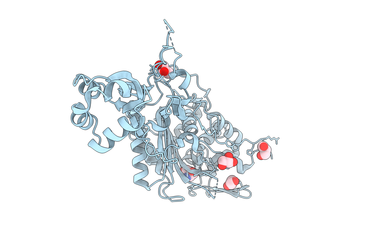 |
Organism: Pseudomonas aeruginosa (strain atcc 15692 / dsm 22644 / cip 104116 / jcm 14847 / lmg 12228 / 1c / prs 101 / pao1)
Method: X-RAY DIFFRACTION Resolution:2.16 Å Release Date: 2021-08-04 Classification: MEMBRANE PROTEIN Ligands: GOL, TRS |

