Planned Maintenance: Some services may turn out to be unavailable from 15th January, 2026 to 16th January, 2026. We apologize for the inconvenience!

Planned Maintenance: Some services may turn out to be unavailable from 15th January, 2026 to 16th January, 2026. We apologize for the inconvenience!
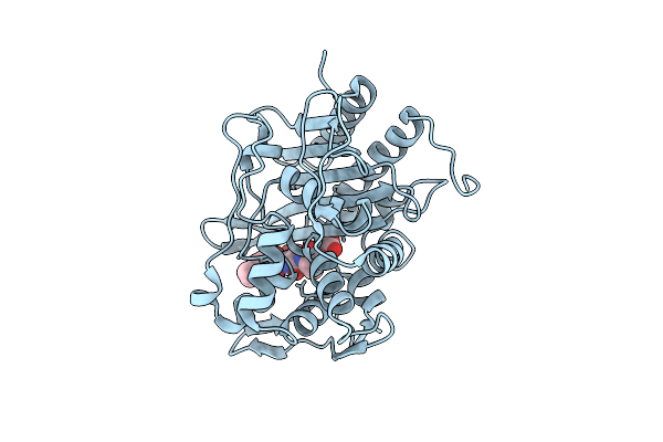 |
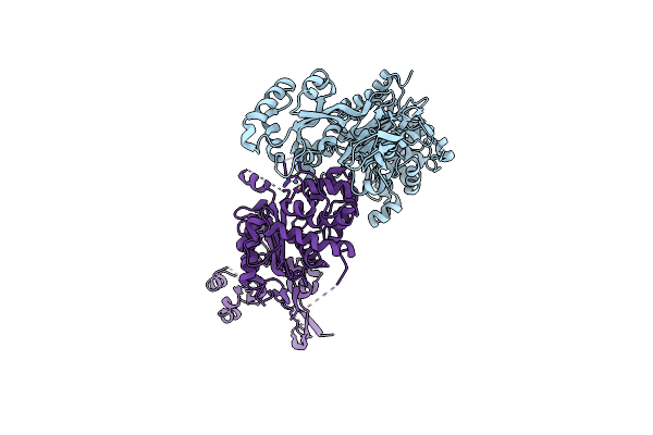
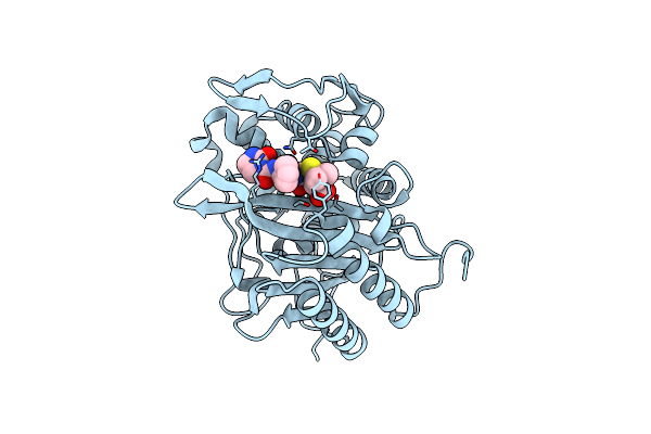
Crystal Structure Of The Transpeptidase Domain Of Pbp2 From The Neisseria Gonorrhoeae Cephalosporin Decreased Susceptibility Strain 35/02 In Complex With Boronate Inhibitor Vnrx-6884
Organism: Neisseria gonorrhoeae 35/02
Method: X-RAY DIFFRACTION Resolution:1.89 Å Release Date: 2025-01-22 Classification: LIGASE Ligands: A1BJC |
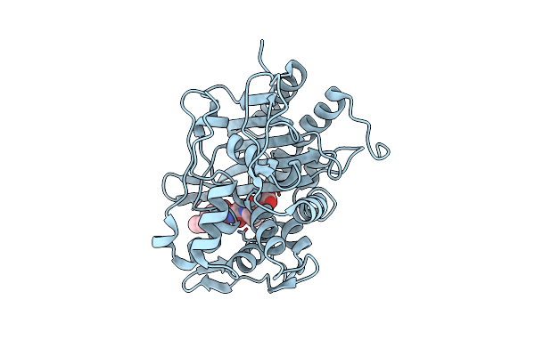 |
Crystal Structure Of The Transpeptidase Domain Of Pbp2 From The Neisseria Gonorrhoeae Cephalosporin Decreased Susceptibility Strain 35/02 In Complex With Boronate Inhibitor Vnrx-6752
Organism: Neisseria gonorrhoeae 35/02
Method: X-RAY DIFFRACTION Resolution:2.61 Å Release Date: 2025-01-22 Classification: LIGASE Ligands: A1BJB |
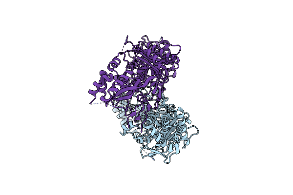 |
Organism: Acinetobacter baumannii
Method: X-RAY DIFFRACTION Resolution:3.31 Å Release Date: 2024-12-04 Classification: HYDROLASE |
 |
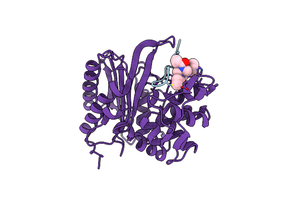
Pseudomonas Aeruginosa Penicillin Binding Protein 3 In Complex With Cefiderocol
Organism: Pseudomonas aeruginosa
Method: X-RAY DIFFRACTION Resolution:1.80 Å Release Date: 2024-10-09 Classification: HYDROLASE Ligands: CAZ |
 |
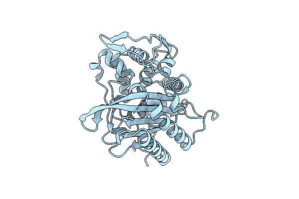
Organism: Pseudomonas aeruginosa
Method: X-RAY DIFFRACTION Resolution:2.11 Å Release Date: 2024-10-09 Classification: HYDROLASE |
 |
Pseudomonas Aeruginosa Penicillin Binding Protein 3 In Complex With Meropenem
Organism: Pseudomonas aeruginosa
Method: X-RAY DIFFRACTION Resolution:2.10 Å Release Date: 2024-10-09 Classification: HYDROLASE Ligands: MER |
 |
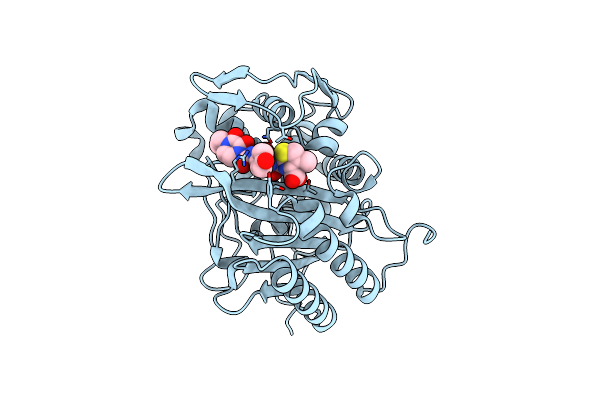
Pseudomonas Aeruginosa Penicillin Binding Protein 3 In Complex With Ceftazidime
Organism: Pseudomonas aeruginosa
Method: X-RAY DIFFRACTION Resolution:1.80 Å Release Date: 2024-10-09 Classification: HYDROLASE Ligands: CAZ, SO4 |
 |
Pseudomonas Aeruginosa Penicillin Binding Protein 3 In Complex With Cefepime
Organism: Pseudomonas aeruginosa
Method: X-RAY DIFFRACTION Resolution:2.70 Å Release Date: 2024-10-09 Classification: HYDROLASE Ligands: CEF |
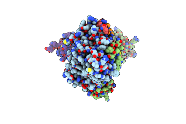 |
Organism: Pseudomonas aeruginosa
Method: ELECTRON MICROSCOPY Release Date: 2024-05-22 Classification: CELL CYCLE |
 |
Organism: Staphylococcaceae bacterium
Method: X-RAY DIFFRACTION Resolution:2.00 Å Release Date: 2024-05-01 Classification: HYDROLASE |
 |
Structure Of The Monofunctional Staphylococcus Aureus Pbp1 In Its Beta-Lactam (Oxacillin) Inhibited Form
Organism: Staphylococcaceae bacterium
Method: X-RAY DIFFRACTION Resolution:2.00 Å Release Date: 2024-05-01 Classification: HYDROLASE/INHIBITOR,ANTIBIOTIC Ligands: 1S6 |
 |
Structure Of The Monofunctional Staphylococcus Aureus Pbp1 In Its Beta-Lactam (Cephalexin) Inhibited Form
Organism: Staphylococcaceae bacterium
Method: X-RAY DIFFRACTION Resolution:2.40 Å Release Date: 2024-05-01 Classification: HYDROLASE/INHIBITOR,ANTIBIOTIC Ligands: 63U |
 |
Structure Of The Monofunctional Staphylococcus Aureus Pbp1 In Its Beta-Lactam (Ertapenem) Inhibited Form
Organism: Staphylococcaceae bacterium
Method: X-RAY DIFFRACTION Resolution:2.30 Å Release Date: 2024-05-01 Classification: HYDROLASE/INHIBITOR,ANTIBIOTIC Ligands: 1RG |
 |
Crystal Structure Of Penicillin-Binding Protein 2 (Pbp2) From Campylobacter Jejuni
Organism: Campylobacter jejuni
Method: X-RAY DIFFRACTION Resolution:2.95 Å Release Date: 2024-04-17 Classification: HYDROLASE Ligands: ZN |
 |
The Structure Of E. Coli Penicillin Binding Protein 3 (Pbp3) In Complex With A Bicyclic Peptide Inhibitor
Organism: Escherichia coli, Synthetic construct
Method: X-RAY DIFFRACTION Release Date: 2024-04-03 Classification: HYDROLASE Ligands: 29N |
 |
Crystal Structure Of The Transpeptidase Domain Of A S310A Mutant Of Pbp2 From Neisseria Gonorrhoeae Strain H041
Organism: Neisseria gonorrhoeae
Method: X-RAY DIFFRACTION Resolution:1.90 Å Release Date: 2024-03-20 Classification: HYDROLASE |
 |
Crystal Structure Of Transpeptidase Domain Of Pbp2 From Neisseria Gonorrhoeae Cephalosporin-Resistant Strain H041 In Complex With Cefoperazone
Organism: Neisseria gonorrhoeae
Method: X-RAY DIFFRACTION Resolution:1.80 Å Release Date: 2024-03-20 Classification: HYDROLASE Ligands: A1ADP |
 |
Crystal Structure Of Transpeptidase Domain Of Pbp2 From Neisseria Gonorrhoeae Cephalosporin-Resistant Strain H041 Acylated By Piperacillin
Organism: Neisseria gonorrhoeae
Method: X-RAY DIFFRACTION Resolution:2.00 Å Release Date: 2024-03-20 Classification: HYDROLASE Ligands: JPP, PEG |
 |
Crystal Structure Of Transpeptidase Domain Of Pbp2 From Neisseria Gonorrhoeae Cephalosporin-Resistant Strain H041 In Complex With Azlocillin
Organism: Neisseria gonorrhoeae
Method: X-RAY DIFFRACTION Resolution:2.40 Å Release Date: 2024-03-20 Classification: HYDROLASE Ligands: 59H |
 |
Crystal Structure Of Enterococcus Faecium Engen25 Penicillin-Binding Protein 5 (Pbp5)
Organism: Enterococcus faecium engen0025
Method: X-RAY DIFFRACTION Resolution:1.90 Å Release Date: 2024-02-07 Classification: PEPTIDE BINDING PROTEIN |