
Search Count: 15
 |
Organism: Homo sapiens, Synthetic construct, Mus musculus
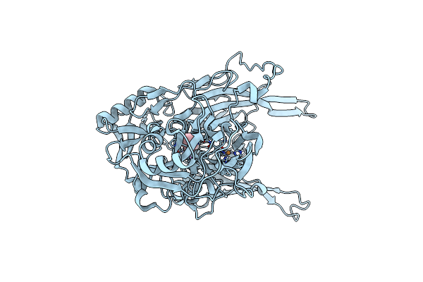
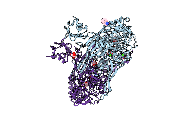
Method: ELECTRON MICROSCOPY Release Date: 2023-12-27 Classification: MEMBRANE PROTEIN Ligands: PEA |
 |
Organism: Homo sapiens, Synthetic construct
Method: ELECTRON MICROSCOPY Release Date: 2023-12-27 Classification: MEMBRANE PROTEIN Ligands: PEA |
 |
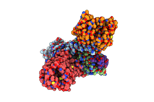
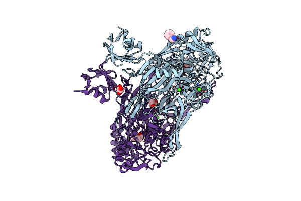
Organism: Homo sapiens, Lama glama
Method: ELECTRON MICROSCOPY Release Date: 2023-11-22 Classification: MEMBRANE PROTEIN Ligands: PEA |
 |
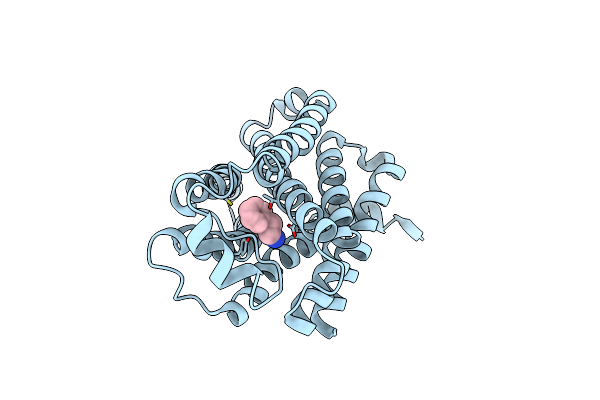
Neutron Structure Of Copper Amine Oxidase From Arthrobacter Globiformis Anaerobically Reduced By Phenylethylamine At Pd 9.0
Organism: Arthrobacter globiformis
Method: X-RAY DIFFRACTION, NEUTRON DIFFRACTION Resolution:1.09 Å, 1.67 Å Release Date: 2023-09-20 Classification: OXIDOREDUCTASE Ligands: CU, NA, PEA |
 |
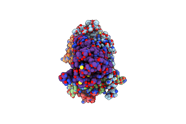
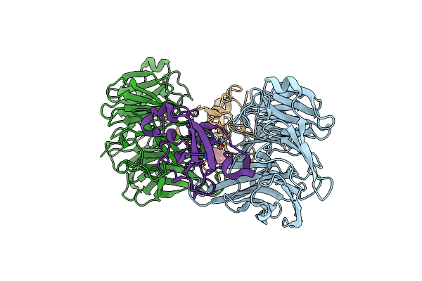
Organism: Mus musculus, Homo sapiens, Escherichia coli bl21(de3)
Method: ELECTRON MICROSCOPY Release Date: 2023-05-31 Classification: MEMBRANE PROTEIN Ligands: PEA |
 |
Organism: Mus musculus
Method: ELECTRON MICROSCOPY Release Date: 2023-05-31 Classification: MEMBRANE PROTEIN Ligands: PEA |
 |
Crystal Structure Of Drosophila Melanogaster Dopamine N-Acetyltransferase Bound To Coa And Phenylethylamine
Organism: Drosophila melanogaster
Method: X-RAY DIFFRACTION Resolution:1.65 Å Release Date: 2017-07-05 Classification: TRANSFERASE Ligands: PEA, COA |
 |
Crystal Structure Of The Complex Of Goat Lactoperoxidase With Phenylethylamine At 2.47 A Resolution
Organism: Capra hircus
Method: X-RAY DIFFRACTION Resolution:2.47 Å Release Date: 2014-01-22 Classification: OXIDOREDUCTASE Ligands: NAG, HEM, CA, IOD, SCN, EDO, PEA |
 |
Crystal Structure Of The Schiff Base Intermediate In The Reductive Half-Reaction Of Aromatic Amine Dehydrogenase (Aadh) With Phenylethylamine.
Organism: Alcaligenes faecalis
Method: X-RAY DIFFRACTION Resolution:1.50 Å Release Date: 2008-04-01 Classification: OXIDOREDUCTASE Ligands: PEA |
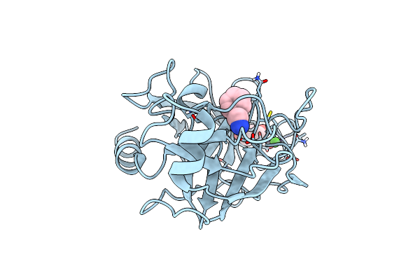 |
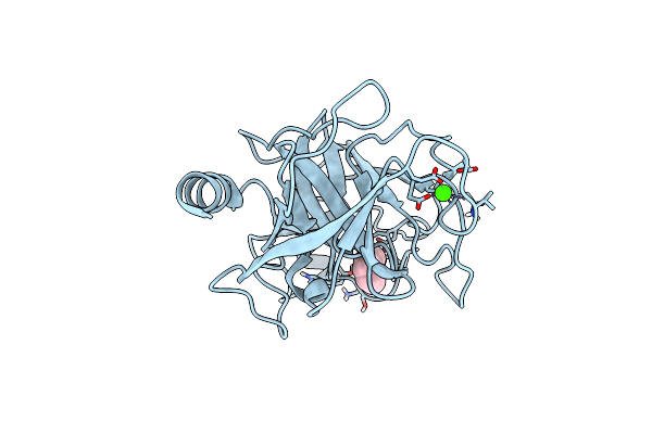
Trypsin Specificity As Elucidated By Lie Calculations, X-Ray Structures And Association Constant Measurements
Organism: Bos taurus
Method: X-RAY DIFFRACTION Resolution:1.15 Å Release Date: 2004-01-15 Classification: HYDROLASE Ligands: PEA, GOL, CA |
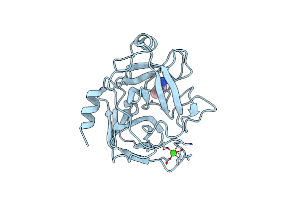 |
Trypsin Specificity As Elucidated By Lie Calculations, X-Ray Structures And Association Constant Measurements
Organism: Salmo salar
Method: X-RAY DIFFRACTION Resolution:1.50 Å Release Date: 2004-01-09 Classification: HYDROLASE Ligands: PEA, CA |
 |
Crystal Structure Of E. Coli Amine Oxidase Anaerobically Reduced With Beta-Phenylethylamine
Organism: Escherichia coli
Method: X-RAY DIFFRACTION Resolution:2.40 Å Release Date: 2000-02-02 Classification: OXIDOREDUCTASE Ligands: CU, CA, HY1, PEA, GOL |
 |
Crystal Structure Of E. Coli Copper-Containing Amine Oxidase Anaerobically Reduced With Beta-Phenylethylamine And Complexed With Nitric Oxide.
Organism: Escherichia coli
Method: X-RAY DIFFRACTION Resolution:2.40 Å Release Date: 2000-02-02 Classification: OXIDOREDUCTASE Ligands: CU, CA, HY1, NO, PEA, GOL |
 |
Crystal Structure Of The Aerobically Freeze Trapped Rate-Determining Catalytic Intermediate Of E. Coli Copper-Containing Amine Oxidase.
Organism: Escherichia coli
Method: X-RAY DIFFRACTION Resolution:2.10 Å Release Date: 2000-02-02 Classification: OXIDOREDUCTASE Ligands: CU, CA, HY1, PEO, PEA, GOL |
 |
Organism: Bos taurus
Method: X-RAY DIFFRACTION Resolution:1.80 Å Release Date: 1994-11-30 Classification: HYDROLASE/hydrolase inhibitor Ligands: CA, PEA |