
Search Count: 55
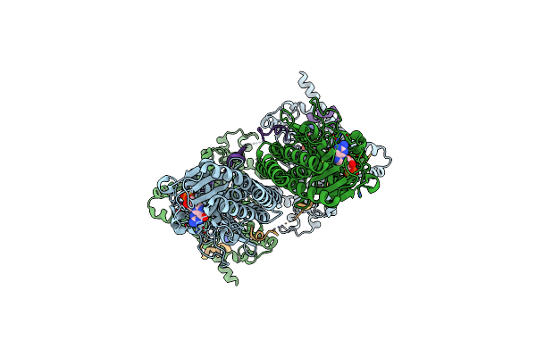 |
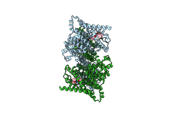
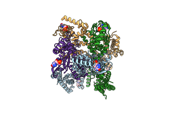
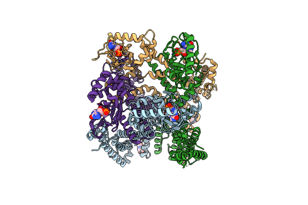
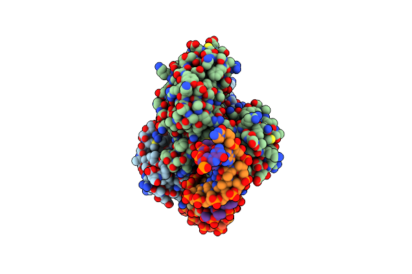
Structure Of Pde6C In Complex With Inhibitory Cone P Gamma In The Presence Of Cgmp
Organism: Homo sapiens
Method: ELECTRON MICROSCOPY Release Date: 2024-12-18 Classification: HYDROLASE Ligands: PCG, ZN, MG, 5GP |
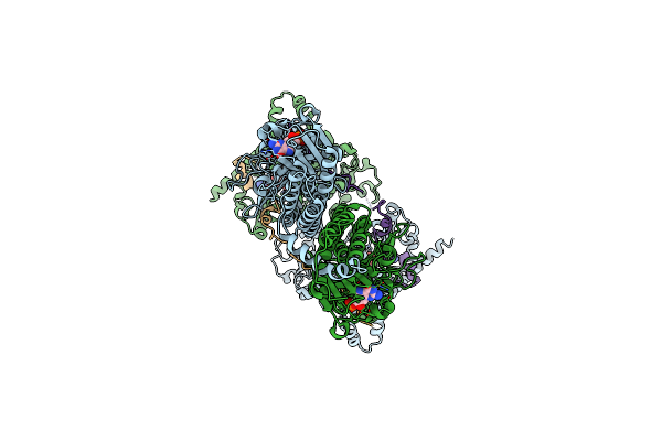 |
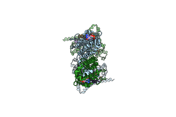
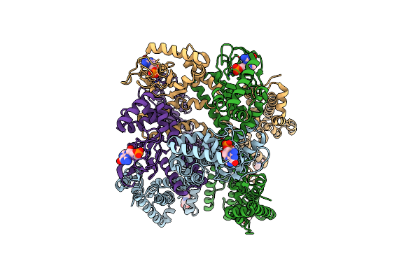
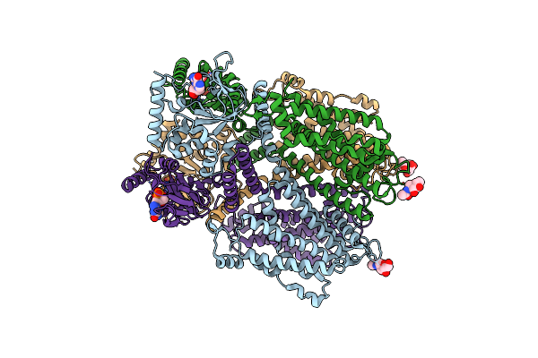
Structure Of Pde6C In Complex With The Rod Inhibitory P Gamma Subunit In The Presence Of Cgmp
Organism: Homo sapiens, Bos taurus
Method: ELECTRON MICROSCOPY Release Date: 2024-12-18 Classification: HYDROLASE Ligands: PCG, ZN, MG, 5GP |
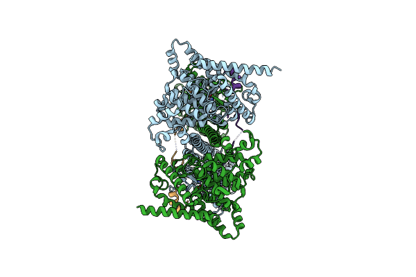 |
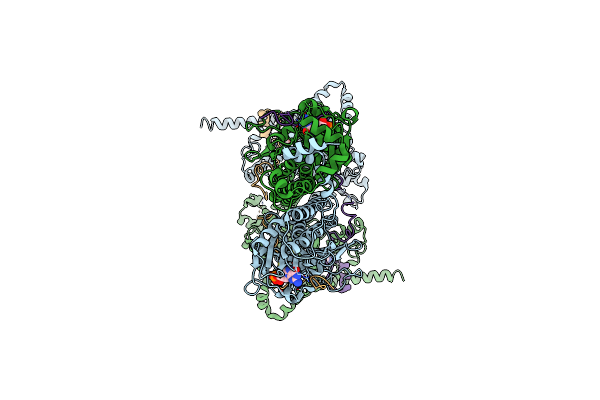
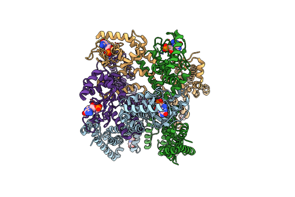
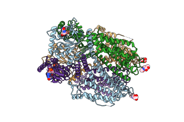
Structure Of Pde6C In Complex With The Rod Inhibitory P Gamma Subunit In The Absence Of Added Cgmp
Organism: Homo sapiens, Mus musculus
Method: ELECTRON MICROSCOPY Release Date: 2024-12-18 Classification: HYDROLASE Ligands: ZN, MG, PCG |
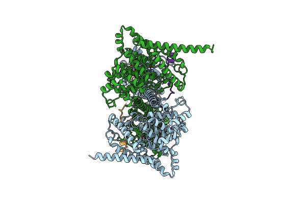 |
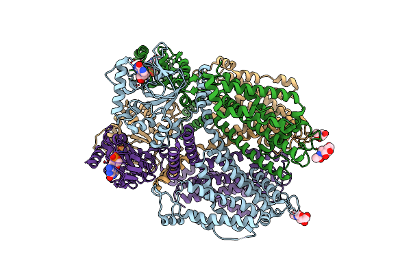
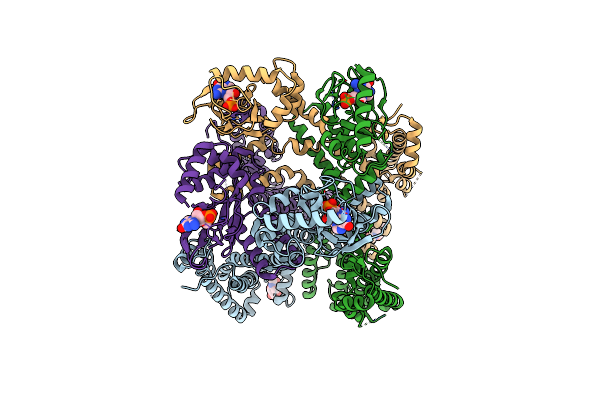
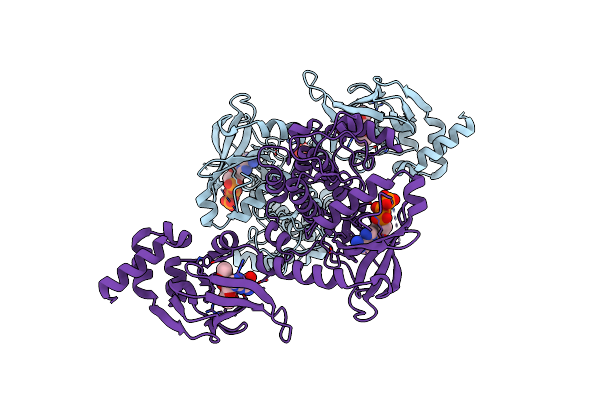
Organism: Bos taurus
Method: ELECTRON MICROSCOPY Release Date: 2024-01-17 Classification: SIGNALING PROTEIN Ligands: PCG, ZN, MG |
 |
Organism: Bos taurus
Method: ELECTRON MICROSCOPY Release Date: 2024-01-17 Classification: SIGNALING PROTEIN/INHIBITOR Ligands: PCG, ZN, MG, ZUD |
 |
Organism: Bos taurus
Method: ELECTRON MICROSCOPY Release Date: 2024-01-17 Classification: SIGNALING PROTEIN/INHIBITOR Ligands: PCG, ZN, MG |
 |
Organism: Bos taurus
Method: ELECTRON MICROSCOPY Release Date: 2024-01-17 Classification: SIGNALING PROTEIN/INHIBITOR Ligands: PCG, MG, ZN, IBM |
 |
Organism: Strongylocentrotus purpuratus
Method: ELECTRON MICROSCOPY Release Date: 2023-11-08 Classification: MEMBRANE PROTEIN Ligands: PCG |
 |
Organism: Strongylocentrotus purpuratus
Method: ELECTRON MICROSCOPY Release Date: 2023-11-08 Classification: MEMBRANE PROTEIN Ligands: PCG |
 |
Cryo-Em Structure Of Cgmp Bound Human Cnga3/Cngb3 Channel In Gdn, Transition State 2
Organism: Homo sapiens
Method: ELECTRON MICROSCOPY Release Date: 2023-08-09 Classification: MEMBRANE PROTEIN Ligands: NAG, PCG |
 |
Cryo-Em Structure Of Cgmp Bound Truncated Human Cnga3/Cngb3 Channel In Lipid Nanodisc, Closed State
Organism: Homo sapiens
Method: ELECTRON MICROSCOPY Release Date: 2023-08-09 Classification: MEMBRANE PROTEIN Ligands: NAG, PCG |
 |
Cryo-Em Structure Of Cgmp Bound Truncated Human Cnga3/Cngb3 Channel In Lipid Nanodisc, Transition State 1
Organism: Homo sapiens
Method: ELECTRON MICROSCOPY Release Date: 2023-08-09 Classification: MEMBRANE PROTEIN Ligands: NAG, PCG |
 |
Cryo-Em Structure Of Cgmp Bound Truncated Human Cnga3/Cngb3 Channel In Lipid Nanodisc, Transition State 2
Organism: Homo sapiens
Method: ELECTRON MICROSCOPY Release Date: 2023-08-09 Classification: MEMBRANE PROTEIN Ligands: NAG, PCG |
 |
Cryo-Em Structure Of Cgmp Bound Truncated Human Cnga3/Cngb3 Channel In Lipid Nanodisc, Pre-Open State
Organism: Homo sapiens
Method: ELECTRON MICROSCOPY Release Date: 2023-08-09 Classification: MEMBRANE PROTEIN Ligands: NAG, PCG |
 |
Cryo-Em Structure Of Cgmp Bound Truncated Human Cnga3/Cngb3 Channel In Lipid Nanodisc, Open State
Organism: Homo sapiens
Method: ELECTRON MICROSCOPY Release Date: 2023-08-09 Classification: MEMBRANE PROTEIN Ligands: NAG, PCG |
 |
Cryo-Em Structure Of Cgmp Bound Closed State Of Human Cnga3/Cngb3 Channel In Gdn
Organism: Homo sapiens
Method: ELECTRON MICROSCOPY Release Date: 2023-08-02 Classification: MEMBRANE PROTEIN Ligands: NAG, PCG |
 |
Cryo-Em Structure Of Cgmp Bound Human Cnga3/Cngb3 Channel In Gdn, Transition State 1
Organism: Homo sapiens
Method: ELECTRON MICROSCOPY Release Date: 2023-08-02 Classification: MEMBRANE PROTEIN Ligands: NAG, PCG |
 |
Organism: Homo sapiens
Method: X-RAY DIFFRACTION Resolution:2.08 Å Release Date: 2023-06-14 Classification: SIGNALING PROTEIN Ligands: PCG, ATP, EDO, CL |
 |
Organism: Sinorhizobium meliloti 1021, Sinorhizobium meliloti
Method: X-RAY DIFFRACTION Release Date: 2022-11-02 Classification: SIGNALING PROTEIN Ligands: PCG |
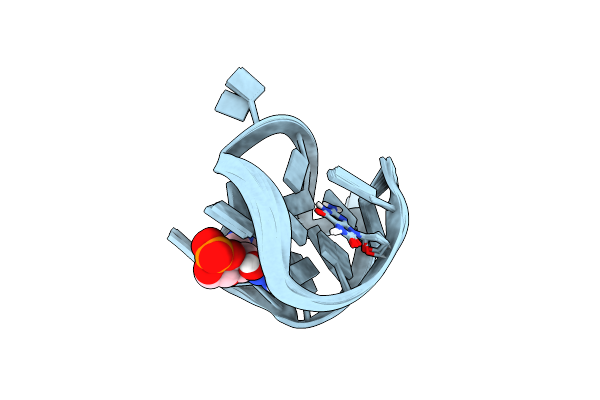 |
Nmr Solution Structure Of A Cgmp Fill-In Vacancy G-Quadruplex Formed In The Oxidized Blm Gene Promoter
Organism: Synthetic construct
Method: SOLUTION NMR Release Date: 2022-05-18 Classification: DNA Ligands: PCG |