
Search Count: 5
 |
Organism: Rhizobium meliloti (strain 1021)
Method: X-RAY DIFFRACTION Resolution:2.80 Å Release Date: 2019-04-17 Classification: MEMBRANE PROTEIN Ligands: TRS, PBE |
 |
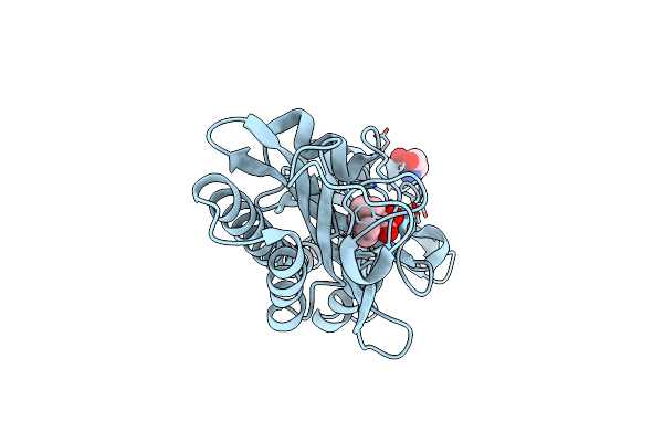
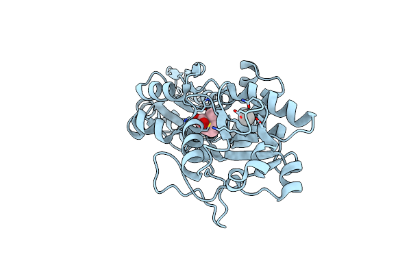
Crystal Structure Of An Enolase (Mandelate Racemase Subgroup) From Paracococus Denitrificans Pd1222 (Target Nysgrc-012907) With Bound L-Proline Betaine (Substrate)
Organism: Paracoccus denitrificans
Method: X-RAY DIFFRACTION Resolution:1.60 Å Release Date: 2013-03-06 Classification: ISOMERASE Ligands: MG, IOD, GOL, PBE |
 |
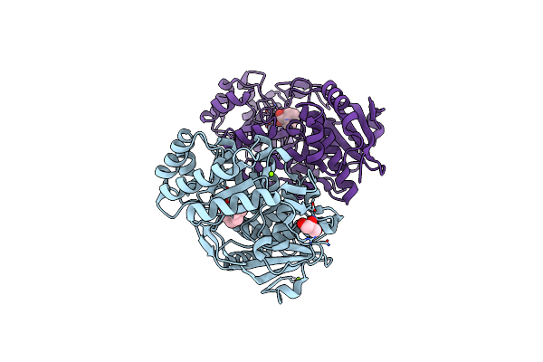
Crystal Structure Of The Binding Protein Opuac In Complex With Proline Betaine
Organism: Bacillus subtilis
Method: X-RAY DIFFRACTION Resolution:2.80 Å Release Date: 2006-03-21 Classification: TRANSPORT PROTEIN Ligands: PBE |
 |
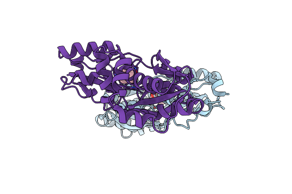
Crystal Structure Of Prox From Archeoglobus Fulgidus In Complex With Proline Betaine
Organism: Archaeoglobus fulgidus
Method: X-RAY DIFFRACTION Resolution:1.90 Å Release Date: 2004-09-14 Classification: PROTEIN BINDING Ligands: ZN, PBE |
 |
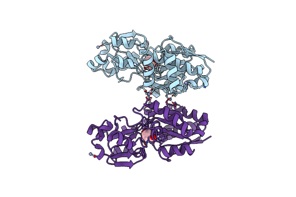
Organism: Escherichia coli
Method: X-RAY DIFFRACTION Resolution:2.05 Å Release Date: 2004-02-24 Classification: PROTEIN BINDING Ligands: UNX, PBE |