
Search Count: 19
 |
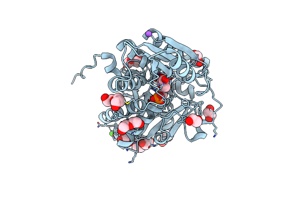
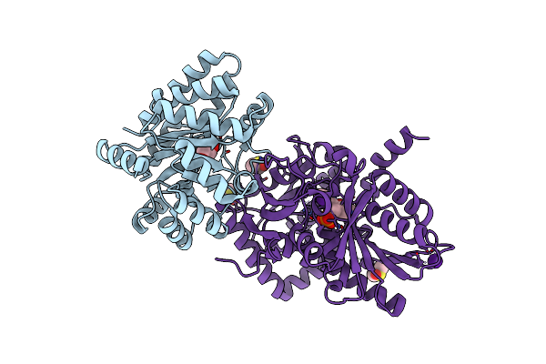
Organism: Pyrenophora teres f. teres 0-1
Method: X-RAY DIFFRACTION Resolution:2.22 Å Release Date: 2023-10-18 Classification: BIOSYNTHETIC PROTEIN Ligands: P1T |
 |
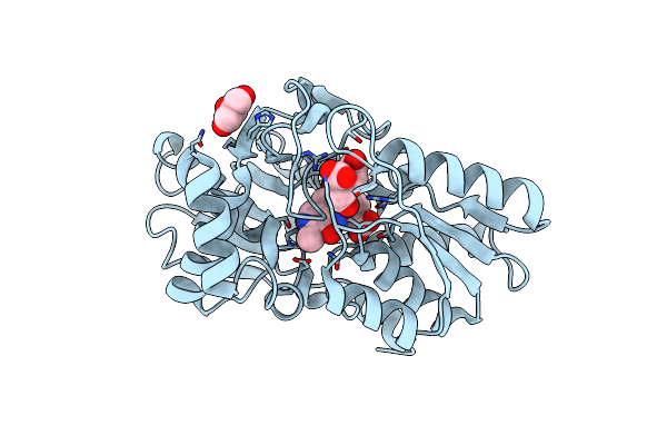
Organism: Saccharothrix mutabilis subsp. capreolus
Method: X-RAY DIFFRACTION Resolution:2.20 Å Release Date: 2023-07-26 Classification: BIOSYNTHETIC PROTEIN Ligands: P1T |
 |
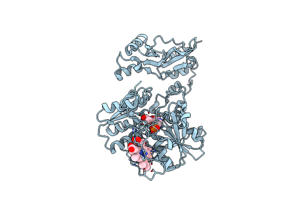
Crystal Structure Of Delta466-491 Cystathionine Beta-Synthase From Toxoplasma Gondii With L-Cysteine
Organism: Toxoplasma gondii (strain atcc 50611 / me49)
Method: X-RAY DIFFRACTION Resolution:3.50 Å Release Date: 2021-07-28 Classification: CYTOSOLIC PROTEIN Ligands: P1T |
 |
Crystal Structure Of Delta466-491 Cystathionine Beta-Synthase From Toxoplasma Gondii With O-Acetylserine
Organism: Toxoplasma gondii me49
Method: X-RAY DIFFRACTION Resolution:3.05 Å Release Date: 2021-06-02 Classification: CYTOSOLIC PROTEIN Ligands: P1T |
 |
Crystal Structure Of Delta466-491 Cystathionine Beta-Synthase From Toxoplasma Gondii With L-Cystathionine
Organism: Toxoplasma gondii (strain atcc 50611 / me49)
Method: X-RAY DIFFRACTION Resolution:3.60 Å Release Date: 2021-02-24 Classification: CYTOSOLIC PROTEIN Ligands: P1T |
 |
Crystal Structure Of Delta466-491 Cystathionine Beta-Synthase From Toxoplasma Gondii With L-Serine
Organism: Toxoplasma gondii me49
Method: X-RAY DIFFRACTION Resolution:3.15 Å Release Date: 2021-02-10 Classification: CYTOSOLIC PROTEIN Ligands: P1T |
 |
Crystal Structure Of Tryptophan Synthase From M. Tuberculosis - Aminoacrylate- And Gsk1-Bound Form
Organism: Mycobacterium tuberculosis (strain atcc 25618 / h37rv)
Method: X-RAY DIFFRACTION Resolution:2.41 Å Release Date: 2020-09-30 Classification: LYASE/Lyase Inhibitor Ligands: FMT, MLI, PZJ, P1T, K, EDO, PEG, ACT, SO4, NA |
 |
Crystal Structure Of Tryptophan Synthase From M. Tuberculosis - Aminoacrylate- And Gsk2-Bound Form
Organism: Mycobacterium tuberculosis (strain atcc 25618 / h37rv)
Method: X-RAY DIFFRACTION Resolution:2.40 Å Release Date: 2020-09-02 Classification: Lyase/Lyase Inhibitor Ligands: FMT, EDO, ACT, MLI, PZV, P1T, PEG, K, EPE, ALA, PGE, NA |
 |
Crystal Structure Of Tryptophan Synthase From M. Tuberculosis - Aminoacrylate- And Brd6309-Bound Form
Organism: Mycobacterium tuberculosis (strain atcc 25618 / h37rv)
Method: X-RAY DIFFRACTION Resolution:2.78 Å Release Date: 2019-09-25 Classification: Lyase/Lyase Inhibitor Ligands: FMT, MLA, P1T, H9V, CS, ACT, PGE, EDO |
 |
Crystal Structure Of Human Cystathionine Gamma Lyase With S-3-Carboxpropyl-L-Cysteine
Organism: Homo sapiens
Method: X-RAY DIFFRACTION Resolution:2.50 Å Release Date: 2019-06-12 Classification: LYASE Ligands: P1T |
 |
Crystal Structure Of Tryptophan Synthase From M. Tuberculosis - Aminoacrylate- And Brd0059-Bound Form
Organism: Mycobacterium tuberculosis (strain atcc 25618 / h37rv)
Method: X-RAY DIFFRACTION Resolution:2.69 Å Release Date: 2018-07-11 Classification: LYASE/LYASE INHIBITOR Ligands: MLI, FMT, P1T, CS, HDJ, ACT, MLA, EDO, PGE |
 |
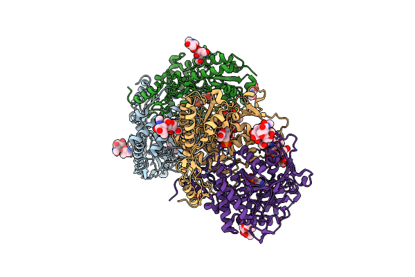
Crystal Structures Of Cystathionine Beta-Synthase From Saccharomyces Cerevisiae: The Structure Of The Plp-Aminoacrylate Intermediate
Organism: Saccharomyces cerevisiae
Method: X-RAY DIFFRACTION Resolution:1.37 Å Release Date: 2018-04-25 Classification: LYASE Ligands: P1T, CA, NA, CL, PEG, PGE, EDO |
 |
Structure Of Mycobacterium Tuberculosis Tryptophan Synthase In Space Group F222
Organism: Mycobacterium tuberculosis (strain atcc 25618 / h37rv)
Method: X-RAY DIFFRACTION Resolution:4.00 Å Release Date: 2017-07-12 Classification: ELECTRON TRANSPORT Ligands: P1T |
 |
Crystal Structure Of Tryptophan Synthase From M. Tuberculosis - Aminoacrylate-Bound Form
Organism: Mycobacterium tuberculosis (strain atcc 25618 / h37rv)
Method: X-RAY DIFFRACTION Resolution:2.40 Å Release Date: 2017-05-31 Classification: LYASE Ligands: MLI, FMT, P1T, CS |
 |
Crystal Structure Of Tryptophan Synthase From M. Tuberculosis - Aminoacrylate And Brd4592-Bound Form
Organism: Mycobacterium tuberculosis (strain atcc 25618 / h37rv)
Method: X-RAY DIFFRACTION Resolution:2.40 Å Release Date: 2017-05-31 Classification: LYASE Ligands: MLI, FMT, P1T, CS, 79V |
 |
Staphyloferrin B Precursor Biosynthetic Enzyme Sbna Bound To Aminoacrylate Intermediate
Organism: Staphylococcus aureus
Method: X-RAY DIFFRACTION Resolution:1.92 Å Release Date: 2016-02-03 Classification: BIOSYNTHETIC PROTEIN Ligands: FLC, P1T, GOL |
 |
Full Length Structure Of Cystathionine Beta-Synthase From Drosophila In Complex With Aminoacrylate
Organism: Drosophila melanogaster
Method: X-RAY DIFFRACTION Resolution:1.55 Å Release Date: 2010-12-01 Classification: LYASE Ligands: HEM, P1T |
 |
Tryptophan Synthase In Complex With Gp, Alpha-D,L-Glycerol-Phosphate, Cs, Ph6.5 - Alpha Aminoacrylate Form - (Gp)E(A-A)
Organism: Salmonella typhimurium
Method: X-RAY DIFFRACTION Resolution:1.90 Å Release Date: 2007-06-26 Classification: LYASE Ligands: G3P, P1T, CS, DMS |
 |
Organism: Allium sativum
Method: X-RAY DIFFRACTION Resolution:1.40 Å Release Date: 2007-02-06 Classification: LYASE Ligands: NAG, CL, P1T |