
Search Count: 33
 |
Organism: Homo sapiens
Method: X-RAY DIFFRACTION Resolution:2.65 Å Release Date: 2019-02-13 Classification: HYDROLASE Ligands: FNB |
 |
Organism: Homo sapiens
Method: X-RAY DIFFRACTION Resolution:2.43 Å Release Date: 2019-01-02 Classification: HYDROLASE Ligands: CW5, MG, GOL |
 |
X-Ray Crystal Structure Of Pf-M1 In Complex With Inhibitor (6Da) And Catalytic Zinc Ion
Organism: Plasmodium falciparum (isolate fcb1 / columbia)
Method: X-RAY DIFFRACTION Resolution:1.82 Å Release Date: 2018-12-26 Classification: hydrolase/hydrolase inhibitor Ligands: ZN, J0Y, GOL, MG |
 |
X-Ray Crystal Structure Of Pf-M1 In Complex With Inhibitor (6H) And Catalytic Zinc Ion
Organism: Plasmodium falciparum (isolate fcb1 / columbia)
Method: X-RAY DIFFRACTION Resolution:1.35 Å Release Date: 2018-12-26 Classification: hydrolase/hydrolase inhibitor Ligands: ZN, J1G, DMS, GOL, MG |
 |
X-Ray Crystal Structure Of Pf-M1 In Complex With Inhibitor (6I) And Catalytic Zinc Ion
Organism: Plasmodium falciparum (isolate fcb1 / columbia)
Method: X-RAY DIFFRACTION Resolution:1.65 Å Release Date: 2018-12-26 Classification: hydrolase/hydrolase inhibitor Ligands: ZN, J1V, GOL, MG, PO4 |
 |
X-Ray Crystal Structure Of Pf-M1 In Complex With Inhibitor (6J) And Catalytic Zinc Ion
Organism: Plasmodium falciparum (isolate fcb1 / columbia)
Method: X-RAY DIFFRACTION Resolution:1.85 Å Release Date: 2018-12-26 Classification: hydrolase/hydrolase inhibitor Ligands: ZN, J2D, GOL, MG, PO4 |
 |
X-Ray Crystal Structure Of Pf-M17 In Complex With Inhibitor 6I And Regulatory Zinc Ion
Organism: Plasmodium falciparum nf135/5.c10
Method: X-RAY DIFFRACTION Resolution:2.10 Å Release Date: 2018-12-26 Classification: hydrolase/hydrolase inhibitor Ligands: CO3, ZN, J1V, SO4, 1PE, DMS, EDO |
 |
X-Ray Crystal Structure Of Pf-M1 In Complex With Inhibitor (6K) And Catalytic Zinc Ion
Organism: Plasmodium falciparum (isolate fcb1 / columbia)
Method: X-RAY DIFFRACTION Resolution:1.82 Å Release Date: 2018-12-26 Classification: hydrolase/hydrolase inhibitor Ligands: ZN, J4V, PO4, GOL, MG |
 |
X-Ray Crystal Structure Of Pf-M1 In Complex With Inhibitor (6M) And Catalytic Zinc Ion
Organism: Plasmodium falciparum (isolate fcb1 / columbia)
Method: X-RAY DIFFRACTION Resolution:1.58 Å Release Date: 2018-12-26 Classification: hydrolase/hydrolase inhibitor Ligands: ZN, J4S, GOL, PO4, MG |
 |
X-Ray Crystal Structure Of Pf-M1 In Complex With Inhibitor (6O) And Catalytic Zinc Ion
Organism: Plasmodium falciparum (isolate fcb1 / columbia)
Method: X-RAY DIFFRACTION Resolution:1.50 Å Release Date: 2018-12-26 Classification: hydrolase/hydrolase inhibitor Ligands: ZN, J4P, GOL, MG |
 |
X-Ray Crystal Structure Of Pf-M1 In Complex With Inhibitor (6P) And Catalytic Zinc Ion
Organism: Plasmodium falciparum (isolate fcb1 / columbia)
Method: X-RAY DIFFRACTION Resolution:1.50 Å Release Date: 2018-12-26 Classification: hydrolase/hydrolase inhibitor Ligands: ZN, J6A, MG, GOL, DMS, PO4 |
 |
X-Ray Crystal Structure Of Pf-M17 In Complex With Inhibitor (6K) And Regulatory Zinc Ion
Organism: Plasmodium falciparum (isolate hb3)
Method: X-RAY DIFFRACTION Resolution:2.30 Å Release Date: 2018-12-26 Classification: hydrolase/hydrolase inhibitor Ligands: J4V, CO3, ZN, SO4, DMS, 1PE, EDO, 2PE |
 |
Organism: Mycobacterium smegmatis
Method: X-RAY DIFFRACTION Resolution:2.15 Å Release Date: 2013-05-22 Classification: HYDROLASE |
 |
Organism: Mycobacterium smegmatis
Method: X-RAY DIFFRACTION Resolution:1.86 Å Release Date: 2013-05-01 Classification: HYDROLASE |
 |
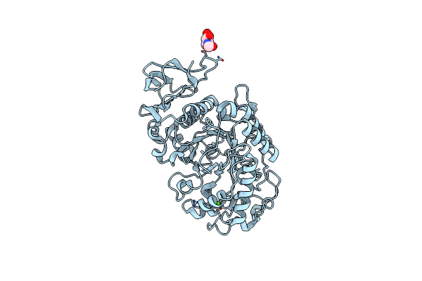
Organism: Homo sapiens
Method: X-RAY DIFFRACTION Resolution:2.00 Å Release Date: 2005-05-24 Classification: HYDROLASE Ligands: NAG, CA |
 |
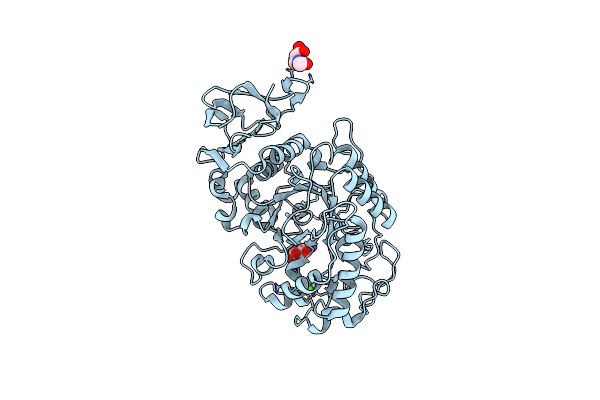
Structure Of The N298S Variant Of Human Pancreatic Alpha-Amylase Complexed With Acarbose
Organism: Homo sapiens
Method: X-RAY DIFFRACTION Resolution:2.00 Å Release Date: 2005-05-24 Classification: HYDROLASE Ligands: NAG, AAO |
 |
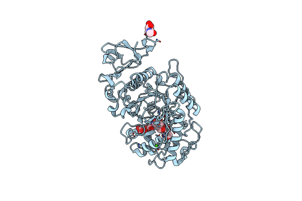
Structure Of The N298S Variant Of Human Pancreatic Alpha-Amylase Complexed With Chloride
Organism: Homo sapiens
Method: X-RAY DIFFRACTION Resolution:2.03 Å Release Date: 2005-05-24 Classification: HYDROLASE Ligands: NAG, CA, CL |
 |
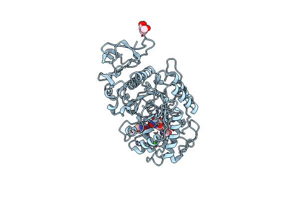
Structure Of The N298S Variant Of Human Pancreatic Alpha-Amylase Complexed With Chloride And Acarbose
Organism: Homo sapiens
Method: X-RAY DIFFRACTION Resolution:2.20 Å Release Date: 2005-05-24 Classification: HYDROLASE Ligands: NAG, ARE, CA, CL |
 |
In Situ Extension As An Approach For Identifying Novel Alpha-Amylase Inhibitors, Structure Containing D-Gluconhydroximo-1,5-Lactam
Organism: Homo sapiens
Method: X-RAY DIFFRACTION Resolution:1.95 Å Release Date: 2004-09-07 Classification: HYDROLASE Ligands: NAG, CA, CL, GOX |
 |
In Situ Extension As An Approach For Identifying Novel Alpha-Amylase Inhibitors, Structure Containing Maltosyl-Alpha (1,4)-D-Gluconhydroximo-1,5-Lactam
Organism: Homo sapiens
Method: X-RAY DIFFRACTION Resolution:1.90 Å Release Date: 2004-09-07 Classification: HYDROLASE Ligands: NAG, LAG, CA, CL, GOX |