
Search Count: 115
 |
Organism: Homo sapiens
Method: ELECTRON MICROSCOPY Release Date: 2025-09-24 Classification: MEMBRANE PROTEIN Ligands: GDP, ZN, NAG, Y01, PLM, NA |
 |
Organism: Homo sapiens
Method: ELECTRON MICROSCOPY Release Date: 2025-09-24 Classification: MEMBRANE PROTEIN Ligands: NAG, GDP, ZN, PLM, ATP |
 |
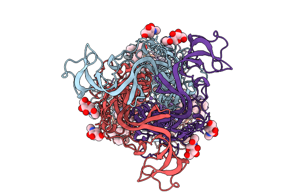
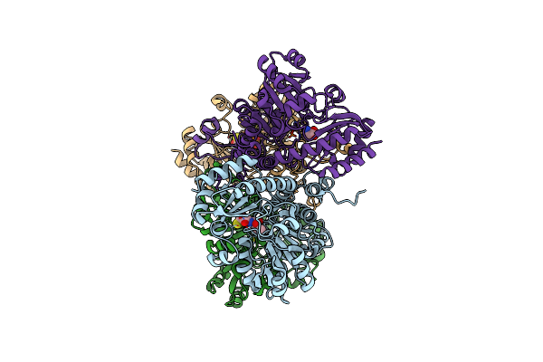
Cryo-Em Structure Of The Human P2X7 Receptor In The Ub-Alt-P30-Bound Inhibited State
Organism: Homo sapiens
Method: ELECTRON MICROSCOPY Release Date: 2025-09-24 Classification: MEMBRANE PROTEIN Ligands: A1BD8, GDP, ZN, NAG, Y01, PLM |
 |
Cryo-Em Structure Of The Human P2X7 Receptor In The Ub-Mbx-46-Bound Inhibited State
Organism: Homo sapiens
Method: ELECTRON MICROSCOPY Release Date: 2025-09-24 Classification: MEMBRANE PROTEIN Ligands: A1BD9, GDP, ZN, NAG, Y01, PLM, NA |
 |
Organism: Mus musculus
Method: ELECTRON MICROSCOPY Release Date: 2025-09-24 Classification: MEMBRANE PROTEIN Ligands: GDP, ZN, NAG, PLM, NA |
 |
Organism: Homo sapiens
Method: X-RAY DIFFRACTION Release Date: 2025-07-30 Classification: NUCLEAR PROTEIN Ligands: A1MAM, EDO, ACT |
 |
Organism: Homo sapiens
Method: ELECTRON MICROSCOPY Release Date: 2025-06-25 Classification: CELL ADHESION Ligands: CA |
 |
Organism: Homo sapiens
Method: ELECTRON MICROSCOPY Release Date: 2025-06-25 Classification: CELL ADHESION |
 |
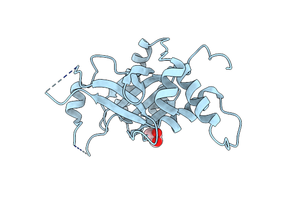
Crystal Structure Of The Mutant Human Ror Gamma Ligand Binding Domain With Jte-151
Organism: Homo sapiens
Method: X-RAY DIFFRACTION Resolution:2.30 Å Release Date: 2024-11-27 Classification: NUCLEAR PROTEIN/INHIBITOR Ligands: YAL |
 |
Organism: Human alphaherpesvirus 1 strain 17
Method: X-RAY DIFFRACTION Resolution:2.32 Å Release Date: 2023-11-29 Classification: VIRAL PROTEIN Ligands: GOL |
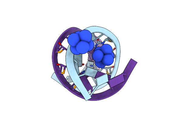 |
Organism: Synthetic construct
Method: X-RAY DIFFRACTION Resolution:1.50 Å Release Date: 2023-05-03 Classification: RNA Ligands: NCO |
 |
Organism: Physarum polycephalum, Gallus gallus
Method: X-RAY DIFFRACTION Resolution:1.15 Å Release Date: 2022-10-26 Classification: CONTRACTILE PROTEIN Ligands: ANP, MG, PO4, EDO, CA |
 |
Organism: Physarum polycephalum, Gallus gallus
Method: X-RAY DIFFRACTION Resolution:1.15 Å Release Date: 2022-10-26 Classification: CONTRACTILE PROTEIN Ligands: ADP, PO4, MG, EDO, CA |
 |
Organism: Physarum polycephalum, Gallus gallus
Method: X-RAY DIFFRACTION Resolution:1.20 Å Release Date: 2022-10-26 Classification: CONTRACTILE PROTEIN Ligands: ADP, MG, EDO, CA |
 |
Organism: Physarum polycephalum, Gallus gallus
Method: X-RAY DIFFRACTION Resolution:2.00 Å Release Date: 2022-10-26 Classification: CONTRACTILE PROTEIN Ligands: ATP, CA, ACT, NA, EDO |
 |
Organism: Physarum polycephalum, Gallus gallus
Method: X-RAY DIFFRACTION Resolution:2.70 Å Release Date: 2022-10-26 Classification: CONTRACTILE PROTEIN Ligands: ADP, CA, ACT, NA, EDO, PO4 |
 |
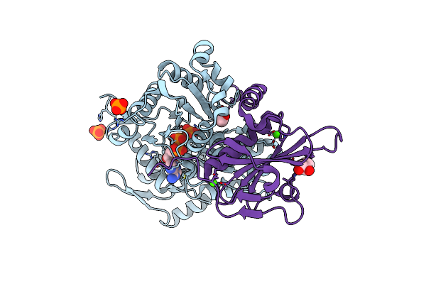
Crystal Structure Of Pseudomonas Putida Methionine Gamma-Lyase Q349S Mutant Ligand-Free Form.
Organism: Pseudomonas putida
Method: X-RAY DIFFRACTION Resolution:2.40 Å Release Date: 2022-04-20 Classification: LYASE |
 |
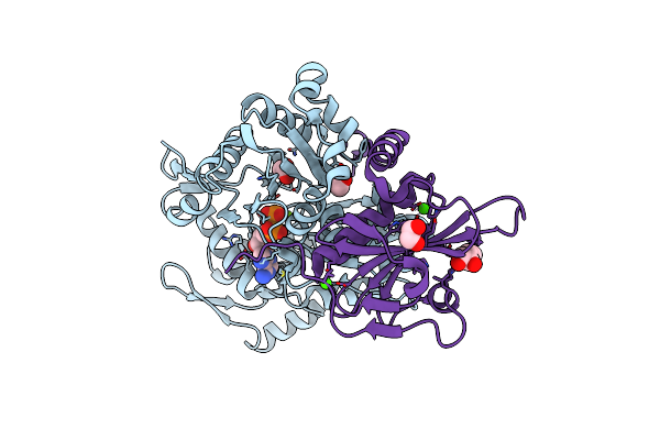
Crystal Structure Of Pseudomonas Putida Methionine Gamma-Lyase Q349S Mutant With L-Methionine Intermediates
Organism: Pseudomonas putida
Method: X-RAY DIFFRACTION Resolution:2.40 Å Release Date: 2022-04-20 Classification: LYASE Ligands: 3LM, MET |
 |
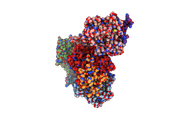
Crystal Structure Of Pseudomonas Putida Methionine Gamma-Lyase Q349S Mutant With L-Homocysteine Intermediates
Organism: Pseudomonas putida
Method: X-RAY DIFFRACTION Release Date: 2022-04-20 Classification: LYASE Ligands: 7XF, HCS |
 |
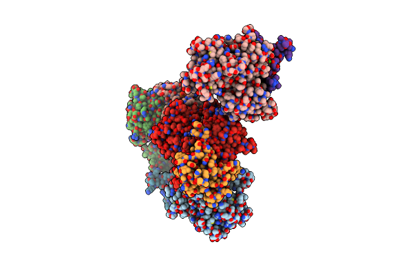
Organism: Severe acute respiratory syndrome coronavirus 2
Method: X-RAY DIFFRACTION Release Date: 2022-03-09 Classification: VIRAL PROTEIN Ligands: SAH, PEG, TRS, ZN |