
Search Count: 164
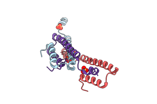 |
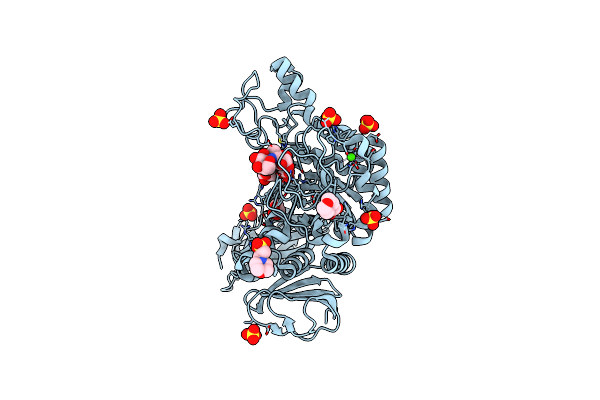
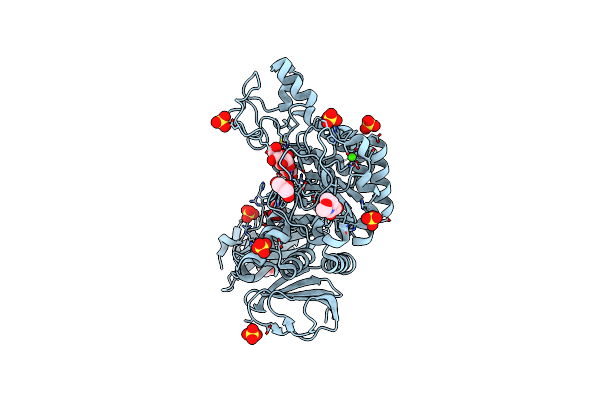
Crystal Structure Of The Semet-Derived C-Terminal Of Viral Responsive Protein 15 (Pmvrp15) From Black Tiger Shrimp Penaeus Monodon
Organism: Penaeus monodon
Method: X-RAY DIFFRACTION Release Date: 2025-10-22 Classification: UNKNOWN FUNCTION Ligands: SO4 |
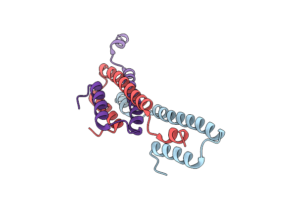 |
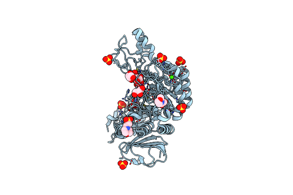
Crystal Structure Of The C-Terminal Of Viral Responsive Protein 15 (Pmvrp15) From Black Tiger Shrimp Penaeus Monodon
Organism: Penaeus monodon
Method: X-RAY DIFFRACTION Release Date: 2025-10-22 Classification: UNKNOWN FUNCTION |
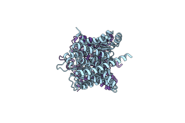 |
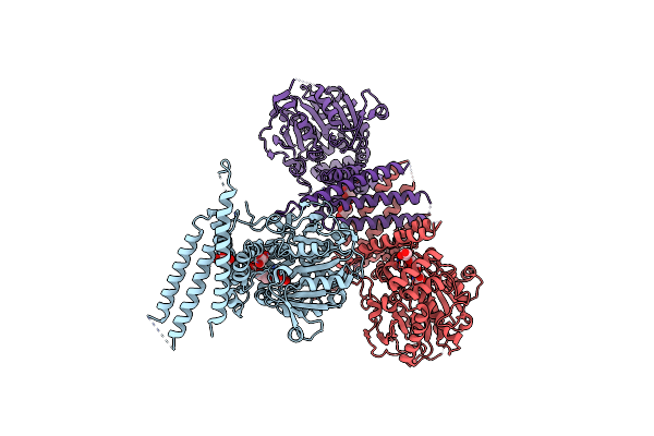
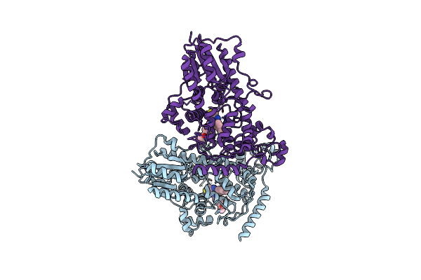
Cryo-Em Structure Of Leminorella Grimontii Gatc In The Presence Of D-Xylose
Organism: Leminorella grimontii
Method: ELECTRON MICROSCOPY Release Date: 2025-10-15 Classification: TRANSPORT PROTEIN |
 |
Organism: Leminorella grimontii
Method: ELECTRON MICROSCOPY Release Date: 2025-10-15 Classification: TRANSPORT PROTEIN |
 |
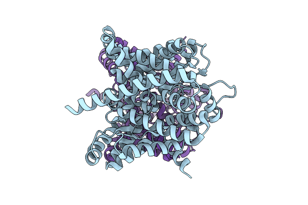
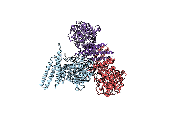
Crystal Structure Of Leminorella Grimontii Gatc In The Presence Of D-Xylose
Organism: Leminorella grimontii
Method: X-RAY DIFFRACTION Release Date: 2025-10-15 Classification: TRANSPORT PROTEIN Ligands: OLC |
 |
Organism: Leminorella grimontii
Method: X-RAY DIFFRACTION Release Date: 2025-10-15 Classification: TRANSPORT PROTEIN Ligands: OLC |
 |
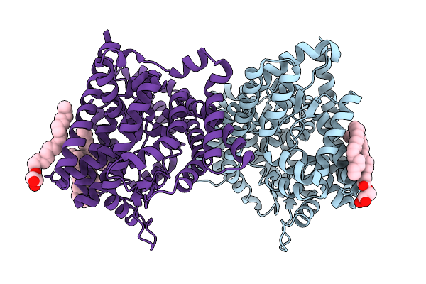
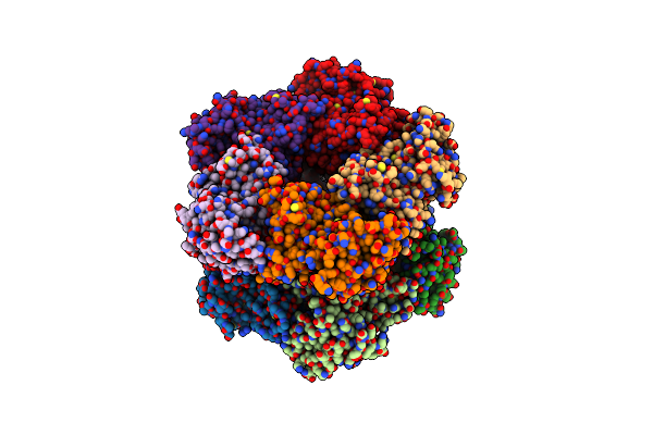
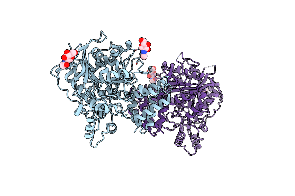
Crystal Structure Of Glycerol-Bound Full-Length Pha Synthase (Phac) From Aeromonas Caviae
Organism: Aeromonas caviae
Method: X-RAY DIFFRACTION Release Date: 2025-05-21 Classification: BIOSYNTHETIC PROTEIN Ligands: GOL |
 |
Organism: Aeromonas caviae
Method: X-RAY DIFFRACTION Release Date: 2025-05-21 Classification: BIOSYNTHETIC PROTEIN |
 |
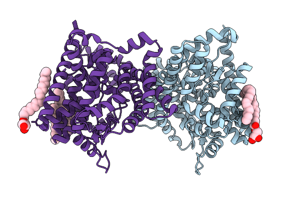
Crystal Structure Of Triethylene Glycol-Bound Full-Length Pha Synthase (Phac) From Aeromonas Caviae
Organism: Aeromonas caviae
Method: X-RAY DIFFRACTION Release Date: 2025-05-21 Classification: BIOSYNTHETIC PROTEIN Ligands: PGE |
 |
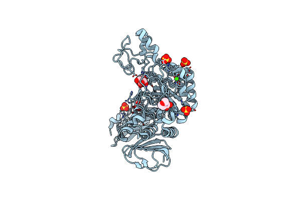
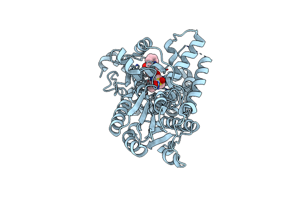
Crystal Structure Of The N-Terminal Degron-Truncated Human Glutamine Synthetase
Organism: Homo sapiens
Method: X-RAY DIFFRACTION Resolution:2.95 Å Release Date: 2021-11-10 Classification: LIGASE |
 |
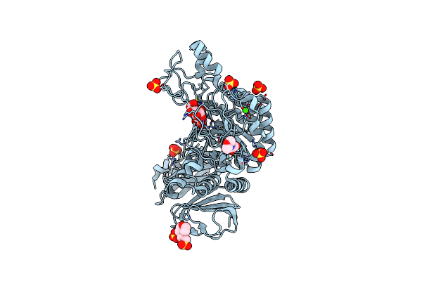
Organism: Weissella cibaria
Method: X-RAY DIFFRACTION Resolution:1.58 Å Release Date: 2021-08-11 Classification: HYDROLASE Ligands: MES, GOL, CA, SO4 |
 |
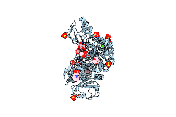
Organism: Weissella confusa
Method: X-RAY DIFFRACTION Resolution:1.36 Å Release Date: 2021-08-11 Classification: HYDROLASE Ligands: CA, SO4, MES, GLC, BGC |
 |
Organism: Weissella cibaria
Method: X-RAY DIFFRACTION Resolution:1.53 Å Release Date: 2021-08-11 Classification: HYDROLASE Ligands: MES, GOL, CA, SO4 |
 |
Organism: Weissella cibaria
Method: X-RAY DIFFRACTION Resolution:1.69 Å Release Date: 2021-08-11 Classification: HYDROLASE Ligands: MES, GOL, CA, SO4 |
 |
Crystal Structure Of Alpha-Glucosidase From Weissella Cibaria Bkk1 In Complex With Maltose
Organism: Weissella cibaria
Method: X-RAY DIFFRACTION Resolution:2.00 Å Release Date: 2021-08-11 Classification: HYDROLASE Ligands: MES, GOL, CA, SO4 |
 |
Organism: Weissella cibaria
Method: X-RAY DIFFRACTION Release Date: 2021-08-11 Classification: HYDROLASE Ligands: MES, GOL, CA, SO4 |
 |
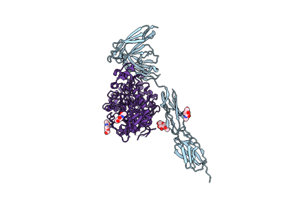
Organism: Mus musculus
Method: X-RAY DIFFRACTION Resolution:2.76 Å Release Date: 2021-02-24 Classification: CELL ADHESION Ligands: NAG |
 |
Organism: Mus musculus
Method: X-RAY DIFFRACTION Resolution:3.85 Å Release Date: 2021-02-24 Classification: HYDROLASE/CELL ADHESION Ligands: NAG |
 |
Organism: Mus musculus
Method: X-RAY DIFFRACTION Resolution:2.35 Å Release Date: 2021-01-27 Classification: CIRCADIAN CLOCK PROTEIN Ligands: GOC |
 |
Organism: Mus musculus
Method: X-RAY DIFFRACTION Resolution:1.91 Å Release Date: 2021-01-27 Classification: CIRCADIAN CLOCK PROTEIN Ligands: GOF |