
Search Count: 5
 |
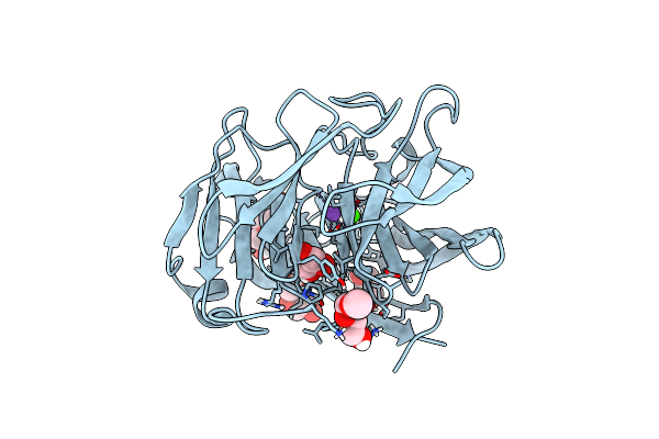
Organism: Homo sapiens
Method: X-RAY DIFFRACTION Resolution:2.15 Å Release Date: 2015-04-01 Classification: PROTEIN BINDING Ligands: CA, P6G, NA |
 |
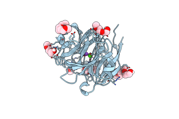
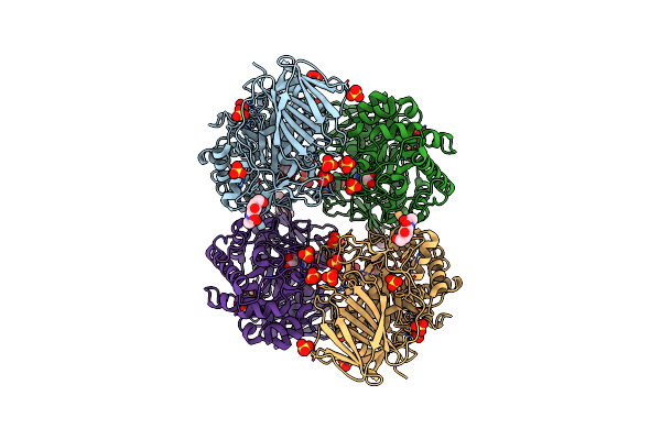
Crystal Structure Of The E396D Snp Variant Of The Myocilin Olfactomedin Domain
Organism: Homo sapiens
Method: X-RAY DIFFRACTION Resolution:1.90 Å Release Date: 2015-04-01 Classification: PROTEIN BINDING Ligands: CA, GOL, PG4, P6G, NA |
 |
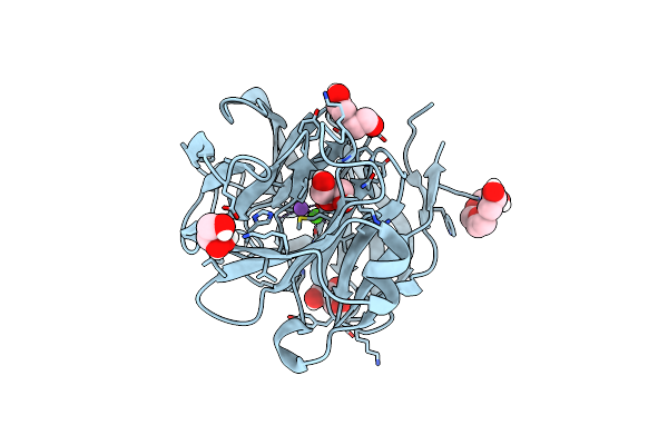
Crystal Structure Of The Selenomthionine Incorporated Myocilin Olfactomedin Domain E396D Variant.
Organism: Homo sapiens
Method: X-RAY DIFFRACTION Resolution:2.09 Å Release Date: 2015-04-01 Classification: PROTEIN BINDING Ligands: CA, P6G, GOL, NA |
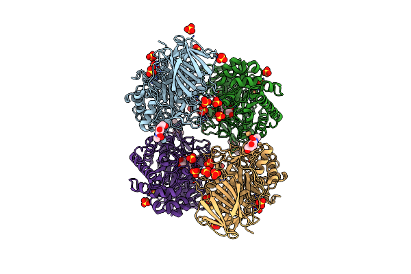 |
The Acid Beta-Glucosidase Active Site Exhibits Plasticity In Binding 3,4,5,6-Tetrahydroxyazepane-Based Inhibitors: Implications For Pharmacological Chaperone Design For Gaucher Disease
Organism: Homo sapiens
Method: X-RAY DIFFRACTION Resolution:2.48 Å Release Date: 2012-03-14 Classification: HYDROLASE/HYDROLASE INHIBITOR Ligands: SO4, NAG, 3RI |
 |
The Acid Beta-Glucosidase Active Site Exhibits Plasticity In Binding 3,4,5,6-Tetrahydroxyazepane-Based Inhibitors: Implications For Pharmacological Chaperone Design For Gaucher Disease
Organism: Homo sapiens
Method: X-RAY DIFFRACTION Resolution:2.40 Å Release Date: 2012-03-14 Classification: HYDROLASE/HYDROLASE INHIBITOR Ligands: SO4, NAG, 3RK |