
Search Count: 15
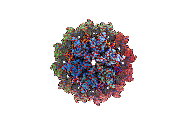 |
Organism: Bacillus subtilis
Method: ELECTRON MICROSCOPY Release Date: 2022-11-30 Classification: VIRAL PROTEIN |
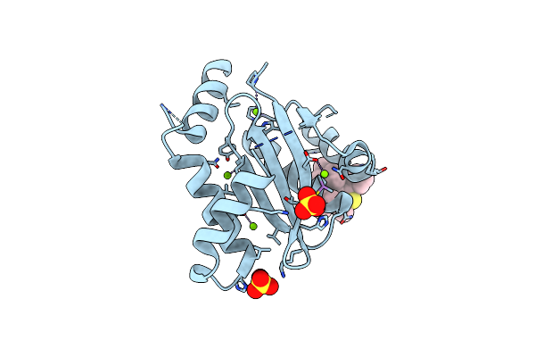 |
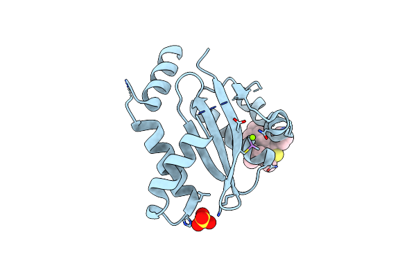
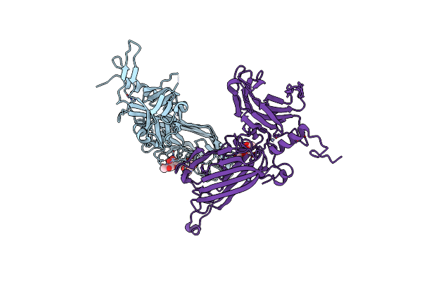
Dissociation Of Biochemical And Antiretroviral Activities Of Integrase-Ledgf Allosteric Inhibitors Revealed By Resistance Of A125 Polymorphic Hiv-1
Organism: Human immunodeficiency virus 1
Method: X-RAY DIFFRACTION Resolution:2.20 Å Release Date: 2018-03-07 Classification: VIRAL PROTEIN Ligands: MG, 9VN, SO4 |
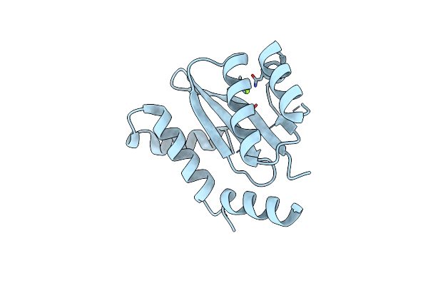 |
Dissociation Of Biochemical And Antiretroviral Activities Of Integrase-Ledgf Allosteric Inhibitors Revealed By Resistance Of A125 Polymorphic Hiv-1
Organism: Human immunodeficiency virus 1
Method: X-RAY DIFFRACTION Resolution:2.30 Å Release Date: 2018-03-07 Classification: VIRAL PROTEIN Ligands: MG |
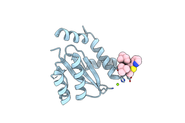 |
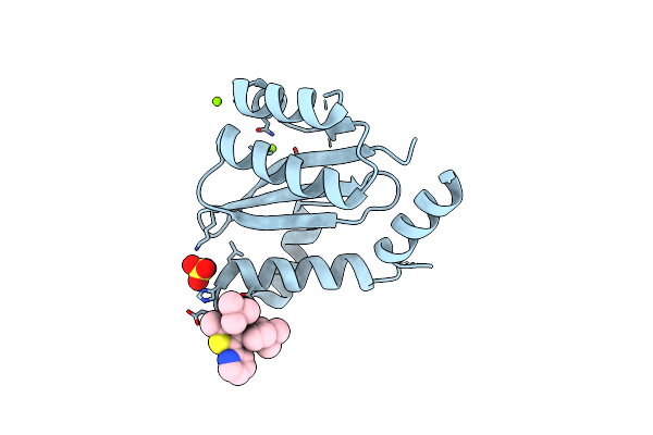
Dissociation Of Biochemical And Antiretroviral Activities Of Integrase-Ledgf Allosteric Inhibitors Revealed By Resistance Of A125 Polymorphic Hiv-1
Organism: Human immunodeficiency virus 1
Method: X-RAY DIFFRACTION Resolution:2.40 Å Release Date: 2018-03-07 Classification: VIRAL PROTEIN Ligands: 9VK, MG |
 |
Dissociation Of Biochemical And Antiretroviral Activities Of Integrase-Ledgf Allosteric Inhibitors Revealed By Resistance Of A125 Polymorphic Hiv-1
Organism: Human immunodeficiency virus 1
Method: X-RAY DIFFRACTION Resolution:2.35 Å Release Date: 2018-03-07 Classification: VIRAL PROTEIN Ligands: MG, 9VN, SO4 |
 |
Dissociation Of Biochemical And Antiretroviral Activities Of Integrase-Ledgf Allosteric Inhibitors Revealed By Resistance Of A125 Polymorphic Hiv-1
Organism: Human immunodeficiency virus 1
Method: X-RAY DIFFRACTION Resolution:2.20 Å Release Date: 2018-03-07 Classification: VIRAL PROTEIN Ligands: MG, 9VK, SO4 |
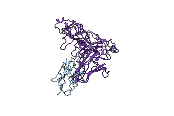 |
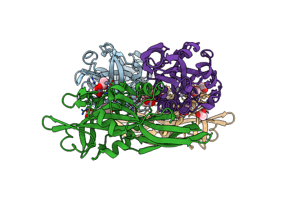
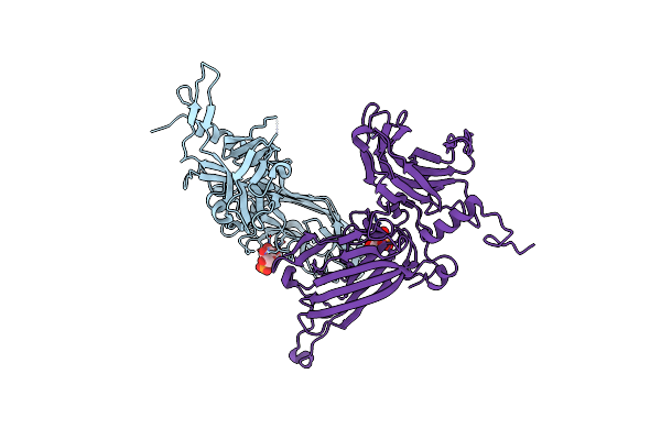
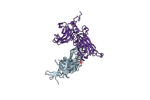
Cryo Electron Microscopy Structure Of Grapevine Fanleaf Virus Complex With Nanobody
Organism: Camelus dromedarius, Grapevine fanleaf virus
Method: ELECTRON MICROSCOPY Resolution:2.80 Å Release Date: 2016-01-20 Classification: VIRUS |
 |
Organism: Pseudomonas entomophila
Method: X-RAY DIFFRACTION Resolution:2.85 Å Release Date: 2015-03-11 Classification: TOXIN Ligands: ZN |
 |
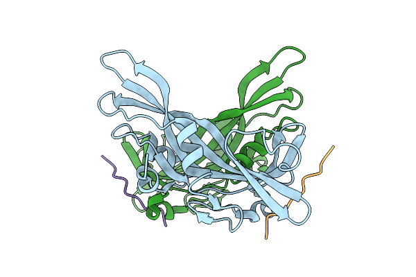
Crystal Structure Of The Monomeric, Cleaved Form Of The Pore-Forming Toxin Monalysin
Organism: Pseudomonas entomophila
Method: X-RAY DIFFRACTION Resolution:1.70 Å Release Date: 2015-03-11 Classification: TOXIN Ligands: HG, ZN, ACT |
 |
Crystal Structure Of The Pore-Forming Toxin Monalysin Mutant Deleted Of The Membrane-Spanning Domain
Organism: Pseudomonas entomophila
Method: X-RAY DIFFRACTION Resolution:2.65 Å Release Date: 2015-03-11 Classification: TOXIN |
 |
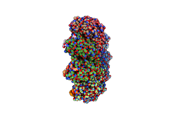
The Cryo-Em Structure Of The Palindromic Dna-Bound Usp-Ecr Nuclear Receptor Reveals An Asymmetric Organization With Allosteric Domain Positioning
Organism: Heliothis virescens, Synthetic construct
Method: ELECTRON MICROSCOPY Resolution:11.60 Å Release Date: 2014-06-25 Classification: NUCLEAR RECEPTOR Ligands: EPH, P1A |
 |
Structure Of The Rbp From Lactococcal Phage 1358 In Complex With 2 Glcnac Molecules
Organism: Lactococcus phage 1358
Method: X-RAY DIFFRACTION Resolution:2.10 Å Release Date: 2014-04-30 Classification: VIRAL PROTEIN Ligands: NDG, NAG, ZN |
 |
Structure Of The Rbp Of Lactococcal Phage 1358 In Complex With Glucose-1-Phosphate
Organism: Lactococcus phage 1358
Method: X-RAY DIFFRACTION Resolution:2.61 Å Release Date: 2014-04-30 Classification: VIRAL PROTEIN Ligands: G1P |
 |
Organism: Lactococcus phage 1358
Method: X-RAY DIFFRACTION Resolution:2.20 Å Release Date: 2014-04-30 Classification: VIRAL PROTEIN Ligands: GOL, ZN |
 |
Organism: Lactococcus phage 1358
Method: X-RAY DIFFRACTION Resolution:1.75 Å Release Date: 2014-04-30 Classification: VIRAL PROTEIN Ligands: CS |