
Search Count: 29
 |
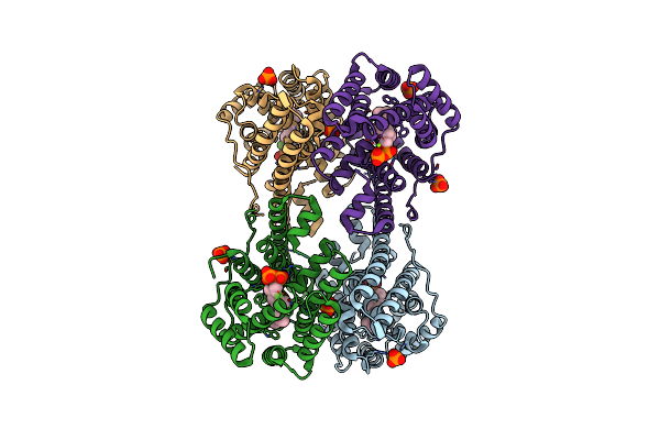
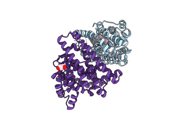
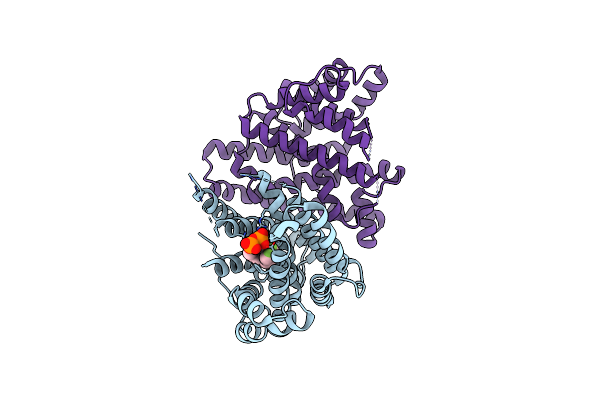
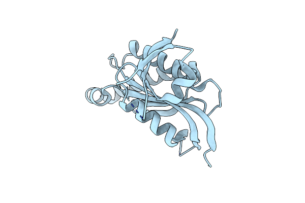
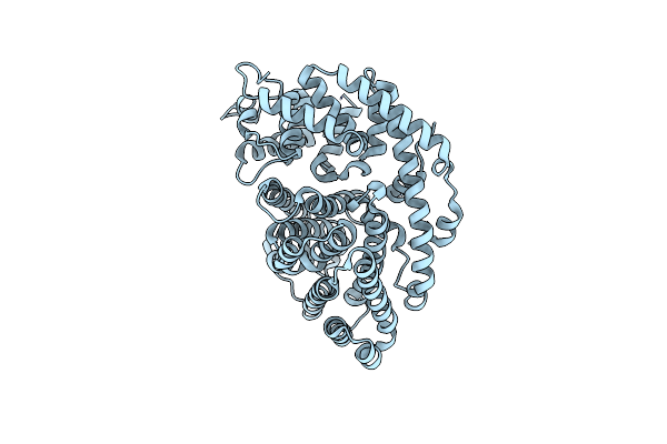
Crystal Structure Of Caryolan-1-Ol Synthase From S. Griseus With Peg Molecule In The Active Site
Organism: Streptomyces griseus
Method: X-RAY DIFFRACTION Resolution:2.33 Å Release Date: 2024-11-06 Classification: METAL BINDING PROTEIN Ligands: PG4, CA, 1PE |
 |
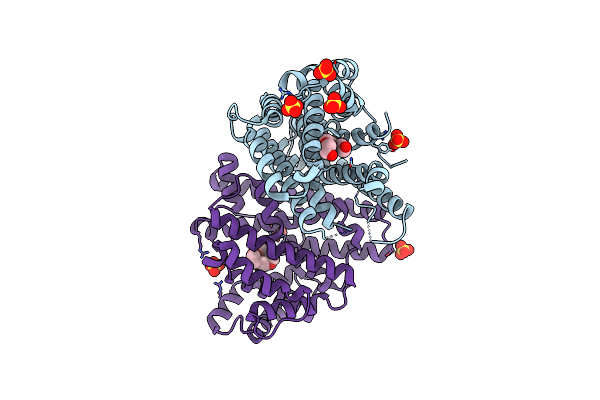
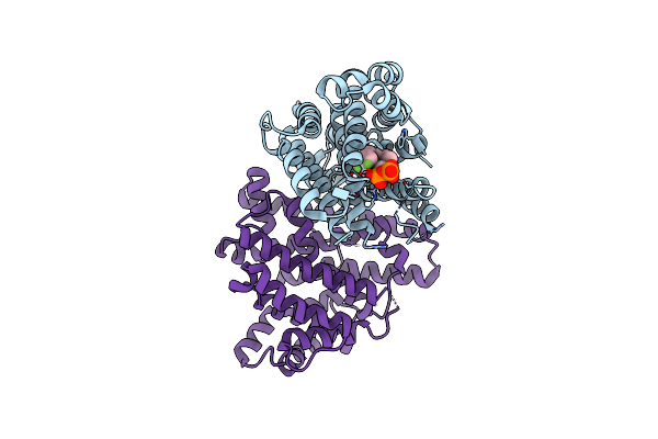
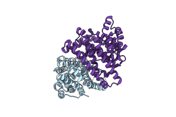
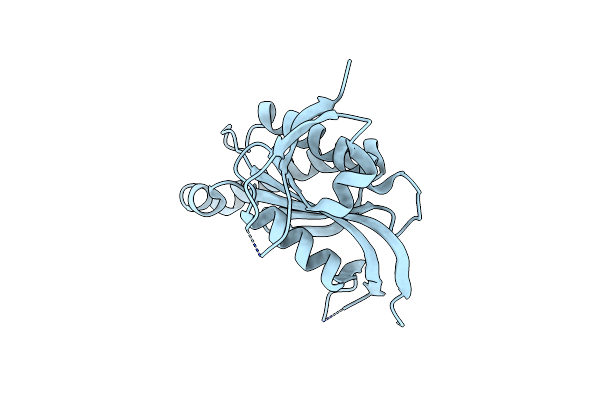
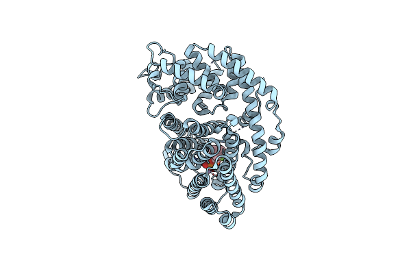
Crystal Structure Of Caryolan-1-Ol Synthase Complexed With 2-Fluorofarnesyl Diphosphate
Organism: Streptomyces griseus
Method: X-RAY DIFFRACTION Resolution:2.65 Å Release Date: 2024-11-06 Classification: METAL BINDING PROTEIN Ligands: FPF, PO4, MG, DPO |
 |
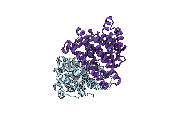
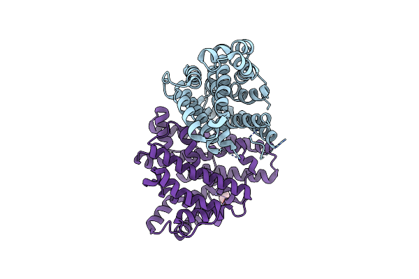
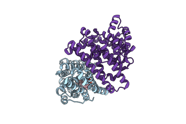
Crystal Structure Of Pentalenene Synthase Variant F76A With Peg Molecule In The Active Site
Organism: Streptomyces exfoliatus
Method: X-RAY DIFFRACTION Resolution:2.50 Å Release Date: 2024-11-06 Classification: METAL BINDING PROTEIN Ligands: PG4, SO4 |
 |
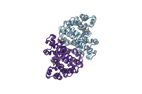
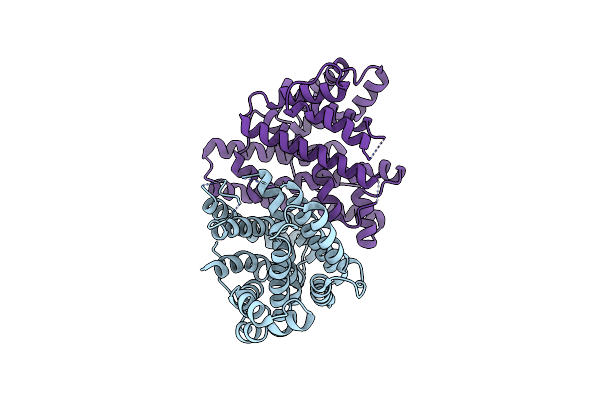
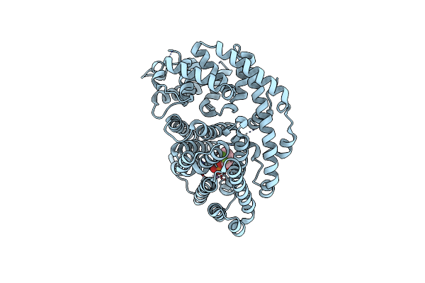
Crystal Structure Of Pentalenene Synthase Variant F76A Complexed With 2-Fluorofarnesyl Diphosphate
Organism: Streptomyces exfoliatus
Method: X-RAY DIFFRACTION Resolution:2.20 Å Release Date: 2024-11-06 Classification: METAL BINDING PROTEIN Ligands: FPF, MG |
 |
Crystal Structure Of Pentalenene Synthase Variant F76A Complexed With 12,13-Difluorofarnesyl Diphosphate
Organism: Streptomyces exfoliatus
Method: X-RAY DIFFRACTION Resolution:2.65 Å Release Date: 2024-11-06 Classification: METAL BINDING PROTEIN Ligands: FDF, MG |
 |
Organism: Streptomyces sp.
Method: X-RAY DIFFRACTION Resolution:1.65 Å Release Date: 2020-08-26 Classification: METAL BINDING PROTEIN, LYASE Ligands: MG |
 |
Crystal Structure Of Pentalenene Synthase Complexed With 12,13-Difluorofarnesyl Diphosphate
Organism: Streptomyces sp.
Method: X-RAY DIFFRACTION Resolution:2.20 Å Release Date: 2020-08-26 Classification: METAL BINDING PROTEIN, LYASE/INHIBITOR Ligands: FDF, GOL, MG |
 |
Organism: Streptomyces sp.
Method: X-RAY DIFFRACTION Resolution:2.40 Å Release Date: 2020-08-26 Classification: METAL BINDING PROTEIN, LYASE Ligands: GOL |
 |
Crystal Structure Of Pentalenene Synthase Mutant F76Y Complexed With 12,13-Difluorofarnesyl Diphosphate
Organism: Streptomyces sp.
Method: X-RAY DIFFRACTION Resolution:2.50 Å Release Date: 2020-08-26 Classification: METAL BINDING PROTEIN, LYASE/INHIBITOR Ligands: FDF |
 |
Organism: Streptomyces sp.
Method: X-RAY DIFFRACTION Resolution:2.30 Å Release Date: 2020-08-26 Classification: METAL BINDING PROTEIN, LYASE |
 |
Crystal Structure Of Pentalenene Synthase Mutant F76W Complexed With 12,13-Difluorofarnesyl Diphosphate
Organism: Streptomyces sp.
Method: X-RAY DIFFRACTION Resolution:2.55 Å Release Date: 2020-08-26 Classification: METAL BINDING PROTEIN, LYASE/INHIBITOR Ligands: FDF |
 |
Organism: Streptomyces sp.
Method: X-RAY DIFFRACTION Resolution:2.35 Å Release Date: 2020-08-26 Classification: METAL BINDING PROTEIN, LYASE |
 |
Crystal Structure Of Pentalenene Synthase Mutant F76H Complexed With 12,13-Difluorofarnesyl Diphosphate
Organism: Streptomyces sp.
Method: X-RAY DIFFRACTION Resolution:2.30 Å Release Date: 2020-08-26 Classification: METAL BINDING PROTEIN, LYASE/INHIBITOR Ligands: FDF |
 |
Crystal Structure Of (+)-Limonene Synthase Complexed With 8,9-Difluorolinalyl Diphosphate
Organism: Citrus sinensis
Method: X-RAY DIFFRACTION Resolution:2.70 Å Release Date: 2019-09-04 Classification: LYASE Ligands: MN, MWG |
 |
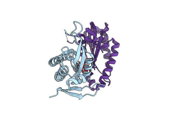
Crystal Structure Of Monomeric Guanylyl Cyclase Domain Of Rhogc Fusion Protein From The Aquatic Fungus Blastocladiella Emersonii
Organism: Blastocladiella emersonii
Method: X-RAY DIFFRACTION Resolution:1.13 Å Release Date: 2017-11-22 Classification: LYASE |
 |
Monomeric Crystal Structure Of The E497/C566D Double Mutant Of The Guanylyl Cyclase Domain Of The Rhogc Fusion Protein From The Aquatic Fungus Blastocladiella Emersonii
Organism: Blastocladiella emersonii
Method: X-RAY DIFFRACTION Resolution:1.40 Å Release Date: 2017-11-22 Classification: LYASE |
 |
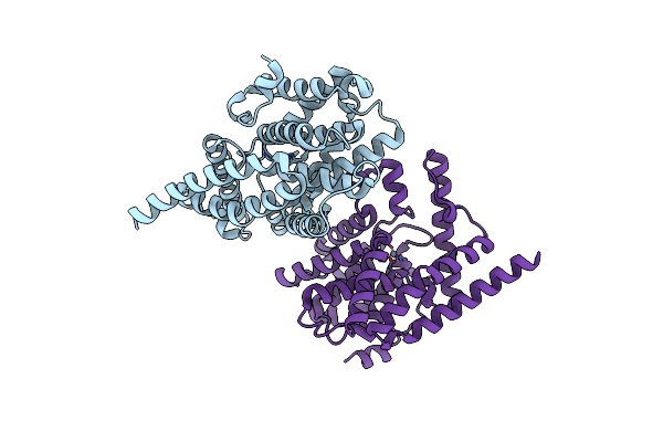
Crystal Structure Of Non-Canonical Dimeric Guanylyl Cyclase Domain Of Rhogc Fusion Protein From The Aquatic Fungus Blastocladiella Emersonii
Organism: Blastocladiella emersonii
Method: X-RAY DIFFRACTION Resolution:1.70 Å Release Date: 2017-11-22 Classification: LYASE Ligands: MN, TLA |
 |
Crystal Structure Of Phosphodiesterase Domain Of Rhopde Fusion Protein From The Choanoflagellate Salpingoeca Rosetta
Organism: Salpingoeca rosetta (strain atcc 50818 / bsb-021)
Method: X-RAY DIFFRACTION Resolution:2.30 Å Release Date: 2017-10-18 Classification: HYDROLASE Ligands: ZN, MG |
 |
Organism: Citrus sinensis
Method: X-RAY DIFFRACTION Resolution:2.30 Å Release Date: 2017-03-22 Classification: LYASE |
 |
Crystal Structure Of (+)-Limonene Synthase Complexed With 2-Fluorogeranyl Diphosphate
Organism: Citrus sinensis
Method: X-RAY DIFFRACTION Resolution:2.40 Å Release Date: 2017-03-22 Classification: LYASE Ligands: MN, 0FV |