
Search Count: 77
 |
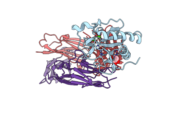
Organism: Homo sapiens, Human gammaherpesvirus 8, Mus musculus
Method: ELECTRON MICROSCOPY Release Date: 2025-09-03 Classification: VIRAL PROTEIN/SIGNALING PROTEIN |
 |
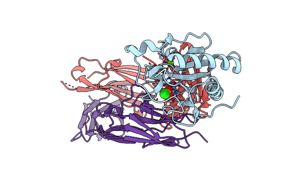
Organism: Human gammaherpesvirus 8
Method: ELECTRON MICROSCOPY Release Date: 2025-08-27 Classification: VIRAL PROTEIN |
 |
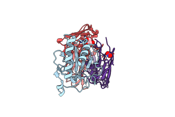
Organism: Human immunodeficiency virus 1, Homo sapiens, Macaca mulatta
Method: ELECTRON MICROSCOPY Release Date: 2025-05-28 Classification: VIRAL PROTEIN/IMMUNE SYSTEM Ligands: NAG |
 |
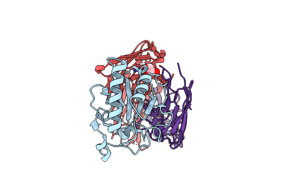
Organism: Human immunodeficiency virus 1, Homo sapiens
Method: ELECTRON MICROSCOPY Release Date: 2025-05-28 Classification: VIRAL PROTEIN/IMMUNE SYSTEM Ligands: NAG |
 |
Crystal Structure Of Sonic Hedgehog In Complex With Antibody 5E1 Mutant H-R102A With Metals
Organism: Homo sapiens
Method: X-RAY DIFFRACTION Resolution:1.89 Å Release Date: 2025-04-16 Classification: IMMUNE SYSTEM Ligands: CL, ZN, CA, GOL |
 |
Crystal Structure Of Sonic Hedgehog In Complex With Antibody 5E1 Mutant L-T56A With Metals
Organism: Homo sapiens
Method: X-RAY DIFFRACTION Resolution:1.80 Å Release Date: 2025-04-16 Classification: IMMUNE SYSTEM Ligands: CA, ZN, CL, SO4 |
 |
Crystal Structure Of Sonic Hedgehog In Complex With Antibody 5E1 Without Metals
Organism: Homo sapiens
Method: X-RAY DIFFRACTION Resolution:1.75 Å Release Date: 2025-04-16 Classification: IMMUNE SYSTEM Ligands: GOL, NO3 |
 |
Crystal Structure Of Sonic Hedgehog In Complex With Antibody 5E1 Mutant H-R102A In The Absence Of Metals
Organism: Homo sapiens
Method: X-RAY DIFFRACTION Resolution:1.90 Å Release Date: 2025-04-09 Classification: IMMUNE SYSTEM Ligands: CL, GOL, IOD |
 |
High-Resolution Structure Of The Siderophore Periplasmic Binding Protein Ftsb From Streptococcus Pyogenes
Organism: Streptococcus pyogenes ssi-1
Method: X-RAY DIFFRACTION Resolution:1.11 Å Release Date: 2024-10-09 Classification: METAL BINDING PROTEIN Ligands: P33, PEG, EDO, P6G, ZN, NA, CL |
 |
Structure Of The Siderophore Periplasmic Binding Protein Ftsb From Streptococcus Pyogenes With Ferrichrome Bound
Organism: Streptococcus pyogenes ssi-1
Method: X-RAY DIFFRACTION Resolution:1.80 Å Release Date: 2024-10-09 Classification: METAL BINDING PROTEIN Ligands: FCE |
 |
Structure Of The Siderophore Periplasmic Binding Protein Ftsb From Streptococcus Pyogenes With Bisucaberin Bound
Organism: Streptococcus pyogenes ssi-1
Method: X-RAY DIFFRACTION Resolution:2.00 Å Release Date: 2024-10-09 Classification: METAL BINDING PROTEIN Ligands: OX8, FE |
 |
High-Resolution Structure Of The Siderophore Periplasmic Binding Protein Ftsb From Streptococcus Pyogenes With Ferrioxamine E Bound
Organism: Streptococcus pyogenes ssi-1
Method: X-RAY DIFFRACTION Resolution:1.12 Å Release Date: 2024-10-09 Classification: METAL BINDING PROTEIN Ligands: 6L0, FE, ZN, NA, GOL, EDO |
 |
High-Resolution Structure Of The Siderophore Periplasmic Binding Protein Ftsb From Streptococcus Pyogenes With Ferrioxamine B
Organism: Streptococcus pyogenes ssi-1
Method: X-RAY DIFFRACTION Resolution:1.15 Å Release Date: 2024-10-09 Classification: METAL BINDING PROTEIN Ligands: 0UE, ZN, EDO, 03S, NA |
 |
High-Resolution Structure Of The Siderophore Periplasmic Binding Protein Ftsb Mutant Y137A From Streptococcus Pyogenes
Organism: Streptococcus pyogenes ssi-1
Method: X-RAY DIFFRACTION Resolution:1.15 Å Release Date: 2024-10-09 Classification: METAL BINDING PROTEIN Ligands: P33, PEG, ZN, SO4, CL, NA |
 |
Structure Of The Siderophore Periplasmic Binding Protein Ftsb Mutant Y137A From Streptococcus Pyogenes With Ferrioxamine E Bound
Organism: Streptococcus pyogenes ssi-1
Method: X-RAY DIFFRACTION Resolution:1.85 Å Release Date: 2024-10-09 Classification: METAL BINDING PROTEIN Ligands: 6L0, FE, GOL |
 |
High-Resolution Structure Of The Siderophore Periplasmic Binding Protein Ftsb From Streptococcus Pyogenes With Ferrioxamine E Bound (Crystal Form 2)
Organism: Streptococcus pyogenes ssi-1
Method: X-RAY DIFFRACTION Resolution:1.95 Å Release Date: 2024-10-09 Classification: METAL BINDING PROTEIN Ligands: 6L0, FE, CL |
 |
Organism: Plasmodium falciparum
Method: X-RAY DIFFRACTION Release Date: 2024-09-04 Classification: DNA BINDING PROTEIN Ligands: SO4 |
 |
Crosslinked 6-Deoxyerythronolide B Synthase (Debs) Module 3 In Complex With Antibody Fragment 1B2: Cis-Oriented 1B2 And Acp
Organism: Saccharopolyspora erythraea, Homo sapiens
Method: ELECTRON MICROSCOPY Release Date: 2024-08-07 Classification: BIOSYNTHETIC PROTEIN |
 |
Crosslinked 6-Deoxyerythronolide B Synthase (Debs) Module 3 In Complex With Antibody Fragment 1B2: Trans-Oriented 1B2 And Acp
Organism: Saccharopolyspora erythraea, Homo sapiens
Method: ELECTRON MICROSCOPY Release Date: 2024-08-07 Classification: BIOSYNTHETIC PROTEIN |
 |
Ks-At Core Of 6-Deoxyerythronolide B Synthase (Debs) Module 3 Crosslinked With Its Translocation Acp Partner Of Module 2
Organism: Saccharopolyspora erythraea
Method: ELECTRON MICROSCOPY Release Date: 2024-07-31 Classification: BIOSYNTHETIC PROTEIN |