
Search Count: 16
 |
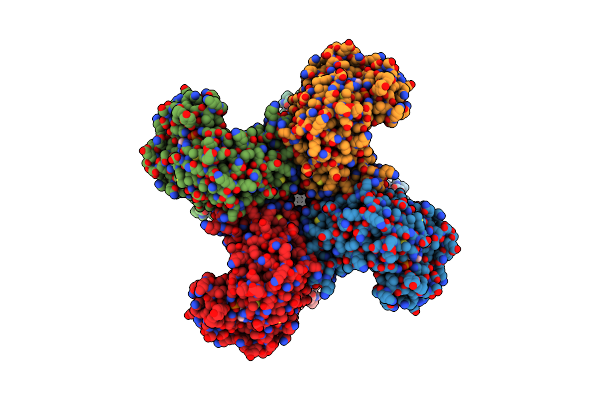
Cryo-Em Structure Of The Human Kca3.1/Calmodulin Channel In Complex With Ca2+ And 1,4-Dihydropyridine (Dhp-103)
Organism: Homo sapiens, Rattus norvegicus
Method: ELECTRON MICROSCOPY Release Date: 2025-04-16 Classification: TRANSPORT PROTEIN Ligands: K, CA |
 |
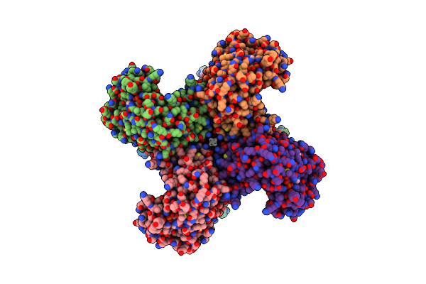
Organism: Homo sapiens
Method: ELECTRON MICROSCOPY Release Date: 2022-02-09 Classification: MEMBRANE PROTEIN Ligands: K, NAP |
 |
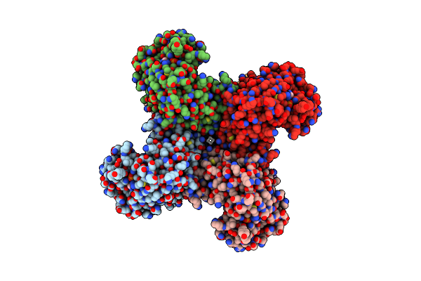
Composite Map Of Human Kv1.3 Channel In Dalazatide-Bound State With Beta Subunits
Organism: Homo sapiens
Method: ELECTRON MICROSCOPY Release Date: 2022-02-09 Classification: MEMBRANE PROTEIN Ligands: NAP, K |
 |
Organism: Homo sapiens
Method: ELECTRON MICROSCOPY Release Date: 2021-06-09 Classification: MEMBRANE PROTEIN Ligands: NAP |
 |
Organism: Homo sapiens
Method: ELECTRON MICROSCOPY Release Date: 2021-06-09 Classification: MEMBRANE PROTEIN Ligands: NAP |
 |
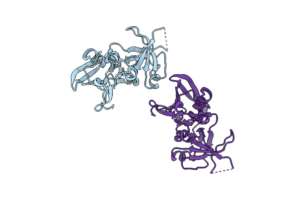
Crystal Structure Of Covid-19 Virus Spike Receptor-Binding Domain Complexed With A Neutralizing Antibody Ct-P59
Organism: Severe acute respiratory syndrome coronavirus 2, Homo sapiens
Method: X-RAY DIFFRACTION Resolution:2.71 Å Release Date: 2021-01-20 Classification: VIRAL PROTEIN Ligands: NI, EDO |
 |
Organism: Vitis vinifera
Method: X-RAY DIFFRACTION Resolution:1.30 Å Release Date: 2020-09-16 Classification: ANTIMICROBIAL PROTEIN |
 |
Organism: Elaeis guineensis
Method: X-RAY DIFFRACTION Resolution:2.10 Å Release Date: 2020-09-09 Classification: PROTEIN BINDING |
 |
Staphylococcal Enterotoxin B (Seb) Mutant S19 - N23A, Y90A, R110A And F177A
Organism: Staphylococcus aureus (strain col)
Method: X-RAY DIFFRACTION Resolution:3.00 Å Release Date: 2017-11-01 Classification: TOXIN |
 |
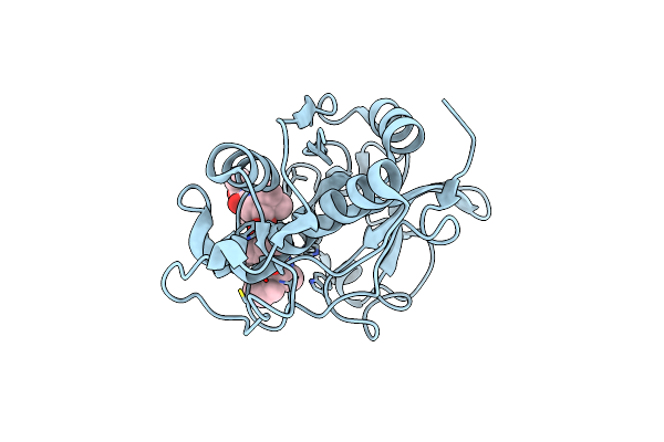
Organism: Homo sapiens
Method: X-RAY DIFFRACTION Resolution:2.40 Å Release Date: 2006-08-08 Classification: HYDROLASE Ligands: CT1 |
 |
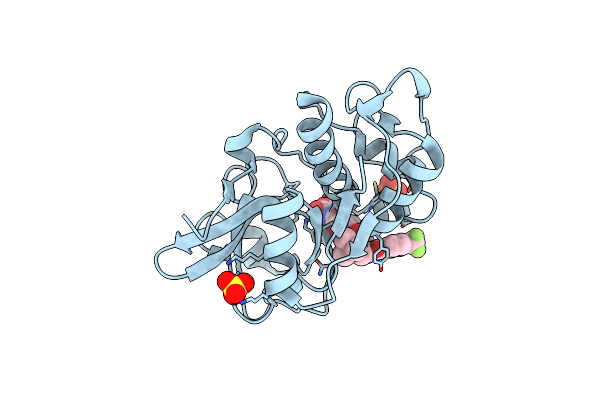
Organism: Homo sapiens
Method: X-RAY DIFFRACTION Resolution:2.30 Å Release Date: 2006-08-08 Classification: HYDROLASE Ligands: SO4, CT2 |
 |
Organism: Dermatophagoides farinae
Method: SOLUTION NMR Release Date: 2006-04-11 Classification: ALLERGEN |
 |
Organism: Homo sapiens
Method: X-RAY DIFFRACTION Resolution:2.00 Å Release Date: 2006-03-21 Classification: HYDROLASE Ligands: 4PR |
 |
Organism: Homo sapiens
Method: X-RAY DIFFRACTION Resolution:2.30 Å Release Date: 2005-07-19 Classification: HYDROLASE Ligands: SO4, 3FC |
 |
Organism: Homo sapiens
Method: X-RAY DIFFRACTION Resolution:1.75 Å Release Date: 2004-09-21 Classification: HYDROLASE Ligands: SO4, FSP |
 |
Organism: Geobacillus stearothermophilus
Method: X-RAY DIFFRACTION Resolution:2.00 Å Release Date: 2002-08-21 Classification: HYDROLASE Ligands: ZN, CA |