Planned Maintenance: Some services may turn out to be unavailable from 15th January, 2026 to 16th January, 2026. We apologize for the inconvenience!

Planned Maintenance: Some services may turn out to be unavailable from 15th January, 2026 to 16th January, 2026. We apologize for the inconvenience!
 |
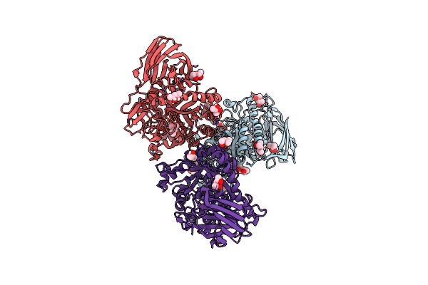
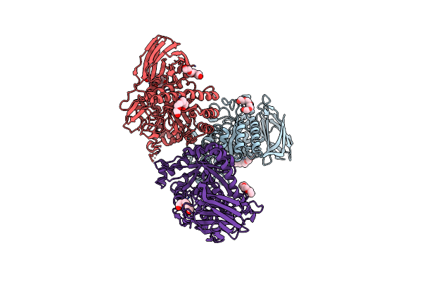
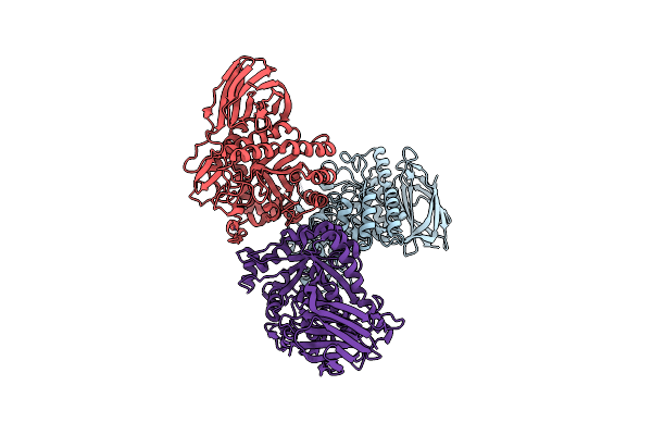
Human Rio1(Kd)-Stha Late Pre-40S Particle, Structural State A (Pre 18S Rrna Cleavage)
Organism: Homo sapiens
Method: ELECTRON MICROSCOPY Release Date: 2021-05-12 Classification: RIBOSOME Ligands: ZN |
 |
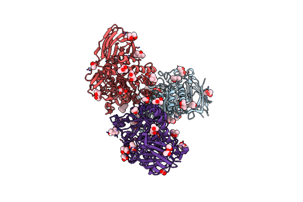
Human Rio1(Kd)-Stha Late Pre-40S Particle, Structural State B (Post 18S Rrna Cleavage)
Organism: Homo sapiens
Method: ELECTRON MICROSCOPY Release Date: 2021-05-12 Classification: RIBOSOME Ligands: MG, ZN, ADP |
 |
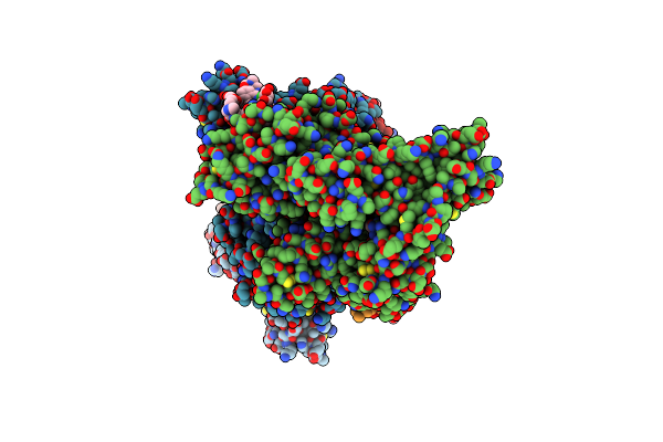
Organism: Thermobacillus xylanilyticus
Method: X-RAY DIFFRACTION Resolution:2.30 Å Release Date: 2021-02-10 Classification: HYDROLASE Ligands: MPD, BTB |
 |
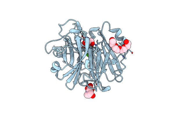
Organism: Thermobacillus xylanilyticus
Method: X-RAY DIFFRACTION Resolution:1.85 Å Release Date: 2021-02-10 Classification: HYDROLASE Ligands: ACT, MPD, BTB, AHR |
 |
Organism: Thermobacillus xylanilyticus
Method: X-RAY DIFFRACTION Resolution:2.80 Å Release Date: 2021-02-10 Classification: HYDROLASE Ligands: 1PE, PG4, CL, PGE |
 |
Organism: Thermobacillus xylanilyticus
Method: X-RAY DIFFRACTION Release Date: 2021-02-10 Classification: HYDROLASE Ligands: AHR, MPD, ACT, BTB |
 |
Organism: Thermobacillus xylanilyticus
Method: X-RAY DIFFRACTION Resolution:3.10 Å Release Date: 2021-02-10 Classification: HYDROLASE |
 |
Organism: Homo sapiens
Method: X-RAY DIFFRACTION Resolution:2.60 Å Release Date: 2020-11-18 Classification: PEPTIDE BINDING PROTEIN Ligands: PO4 |
 |
Organism: Homo sapiens, Synthetic construct
Method: X-RAY DIFFRACTION Resolution:2.80 Å Release Date: 2020-11-18 Classification: PEPTIDE BINDING PROTEIN Ligands: JEF, F8F, M12, MAN, F8X, WOO |
 |
Structure Of Anabaena Sensory Rhodopsin Determined By Solid State Nmr Spectroscopy And Deer
Organism: Nostoc sp. (strain pcc 7120 / sag 25.82 / utex 2576)
Method: SOLID-STATE NMR Release Date: 2017-05-31 Classification: SIGNALING PROTEIN |
 |
Crystal Structure Of The Alpha-L-Arabinofuranosidase Umabf62A From Ustilago Maidys
Organism: Ustilago maydis
Method: X-RAY DIFFRACTION Resolution:1.00 Å Release Date: 2014-01-15 Classification: HYDROLASE Ligands: CA, GOL, TRS, 1PE |
 |
Crystal Structure Of The Alpha-L-Arabinofuranosidase Umabf62A From Ustilago Maydis In Complex With L-Arabinofuranose
Organism: Ustilago maydis
Method: X-RAY DIFFRACTION Resolution:1.20 Å Release Date: 2014-01-15 Classification: HYDROLASE Ligands: CA, FUB, AHR, TRS |
 |
Crystal Structure Of The Alpha-L-Arabinofuranosidase Paabf62A From Podospora Anserina In Complex With Cellotriose
Organism: Podospora anserina
Method: X-RAY DIFFRACTION Resolution:1.80 Å Release Date: 2014-01-15 Classification: HYDROLASE Ligands: CA, TRS, 1PE, EPE |
 |
Crystal Structure Of The Alpha-L-Arabinofuranosidase Paabf62A From Podospora Anserina
Organism: Podospora anserina
Method: X-RAY DIFFRACTION Resolution:1.44 Å Release Date: 2014-01-15 Classification: HYDROLASE Ligands: CA, EPE, TRS, 1PE |
 |
Structure Of A Seleno-Methionyl Derivative Of Wild Type Arabinofuranosidase From Thermobacillus Xylanilyticus
Organism: Thermobacillus xylanilyticus
Method: X-RAY DIFFRACTION Resolution:2.20 Å Release Date: 2008-07-01 Classification: HYDROLASE Ligands: PO4 |
 |
Structure Of An Inactive Mutant Of Arabinofuranosidase From Thermobacillus Xylanilyticus In Complex With A Pentasaccharide
Organism: Thermobacillus xylanilyticus
Method: X-RAY DIFFRACTION Resolution:2.00 Å Release Date: 2008-07-01 Classification: HYDROLASE Ligands: PO4 |
 |
Organism: Phytophthora cryptogea
Method: X-RAY DIFFRACTION Resolution:2.15 Å Release Date: 1999-06-15 Classification: FUNGAL TOXIC ELICITOR Ligands: ERG |