
Search Count: 67
 |
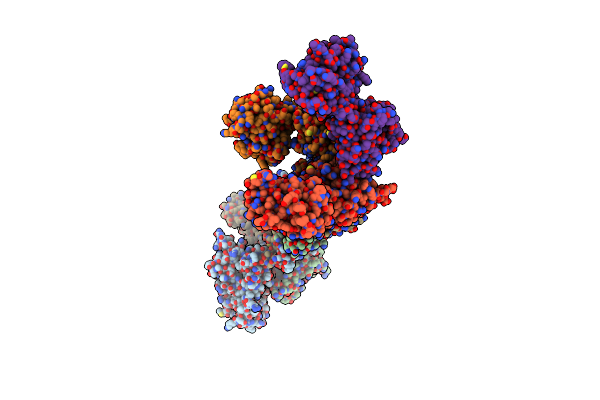
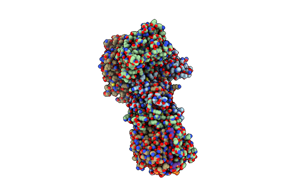
Structure Of Photosynthetic Lh1-Rc Complex From The Purple Bacterium Blastochloris Tepida
Organism: Blastochloris tepida
Method: ELECTRON MICROSCOPY Release Date: 2024-12-18 Classification: PHOTOSYNTHESIS Ligands: HEC, MG, UQ8, DGA, BCB, BPB, CDL, FE, MQ7, NS5, LMT, PGV, NS0 |
 |
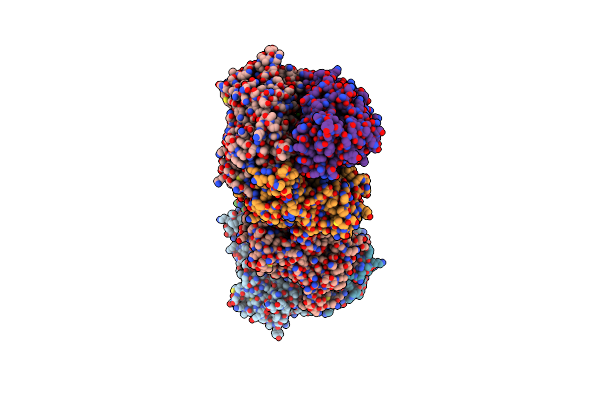
Crystal Structure Of The K87V Mutant Of Cytochrome C' From Shewanella Benthica Db6705
Organism: Shewanella sp. db6705
Method: X-RAY DIFFRACTION Resolution:2.06 Å Release Date: 2023-10-11 Classification: ELECTRON TRANSPORT Ligands: HEC |
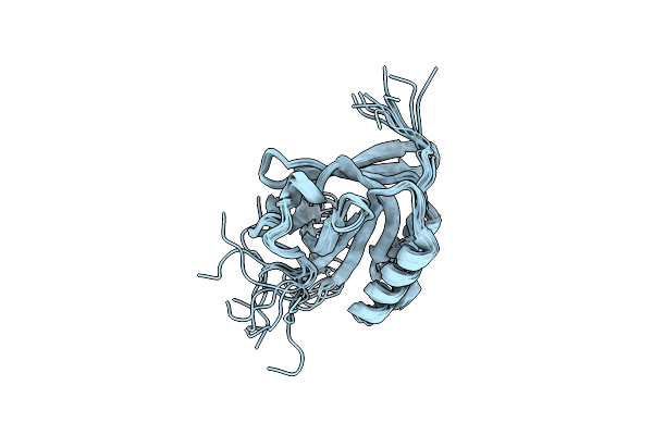 |
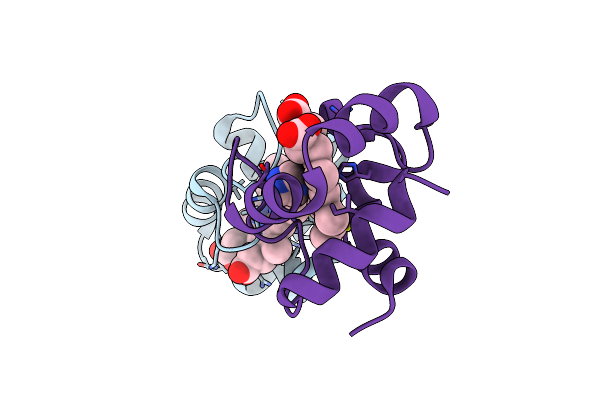
Solution Structure Of The C65A/C167A Mutant Of Human Lipocalin-Type Prostaglandin D Synthase
|
 |
Organism: Vibrio cholerae
Method: X-RAY DIFFRACTION Resolution:2.32 Å Release Date: 2022-11-09 Classification: CELL ADHESION Ligands: SO4 |
 |
Crystal Structure Of Minor Pilin Tcpb From Vibrio Cholerae Complexed With N-Terminal Peptide Fragment Of Tcpf
Organism: Vibrio cholerae
Method: X-RAY DIFFRACTION Resolution:2.30 Å Release Date: 2022-11-09 Classification: CELL ADHESION Ligands: CA, CL, 1PE |
 |
Crystal Structure Of Minor Pilin Tcpb From Vibrio Cholerae Complexed With Secreted Protein Tcpf
Organism: Vibrio cholerae
Method: X-RAY DIFFRACTION Resolution:4.05 Å Release Date: 2022-11-09 Classification: CELL ADHESION |
 |
Crystal Structure Of Beta-Sheet Cytochrome C Prime From Thermus Thermophilus.
Organism: Thermus thermophilus
Method: X-RAY DIFFRACTION Resolution:1.74 Å Release Date: 2022-03-09 Classification: ELECTRON TRANSPORT Ligands: HEC |
 |
Crystal Structure Of Mouse Pedf In Complex With Heterotrimeric Collagen Model Peptide.
Organism: Mus musculus, Homo sapiens
Method: X-RAY DIFFRACTION Resolution:2.48 Å Release Date: 2020-09-02 Classification: SIGNALING PROTEIN Ligands: SO4 |
 |
Organism: Pseudomonas sp.
Method: X-RAY DIFFRACTION Resolution:1.57 Å Release Date: 2020-08-19 Classification: ELECTRON TRANSPORT Ligands: ZN, HEC |
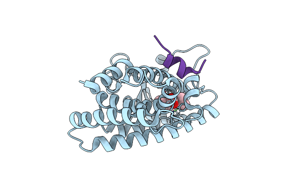 |
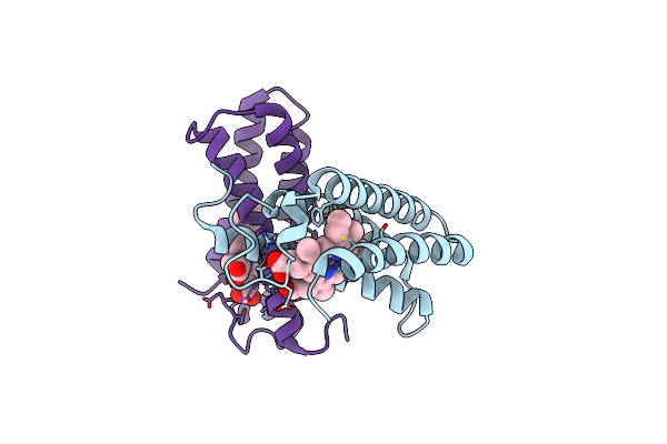
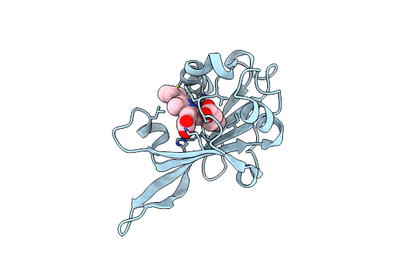
Human Ppar Alpha Ligand Binding Domain In Complex With A Synthetic Agonist (Compound A)
Organism: Homo sapiens
Method: X-RAY DIFFRACTION Resolution:1.95 Å Release Date: 2020-05-20 Classification: TRANSCRIPTION Ligands: T02 |
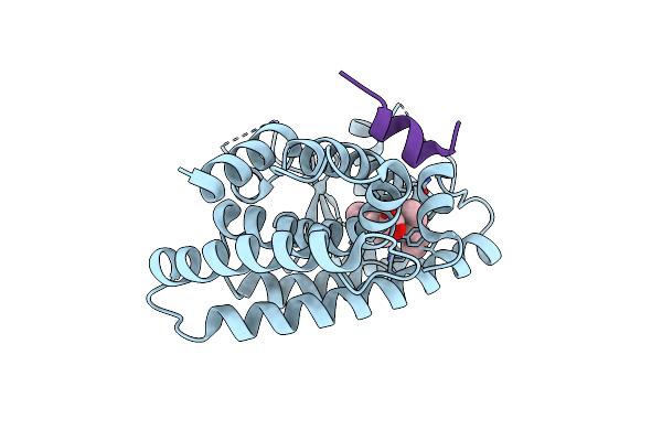 |
Human Ppar Alpha Ligand Binding Domain In Complex With A Synthetic Agonist (Compound B)
Organism: Homo sapiens
Method: X-RAY DIFFRACTION Resolution:2.00 Å Release Date: 2020-05-20 Classification: TRANSCRIPTION Ligands: T06 |
 |
Organism: Shewanella benthica db6705
Method: X-RAY DIFFRACTION Resolution:1.71 Å Release Date: 2019-06-12 Classification: ELECTRON TRANSPORT Ligands: HEC, 1PE |
 |
Organism: Shewanella violacea (strain jcm 10179 / cip 106290 / lmg 19151 / dss12)
Method: X-RAY DIFFRACTION Resolution:2.14 Å Release Date: 2019-06-12 Classification: ELECTRON TRANSPORT Ligands: HEC |
 |
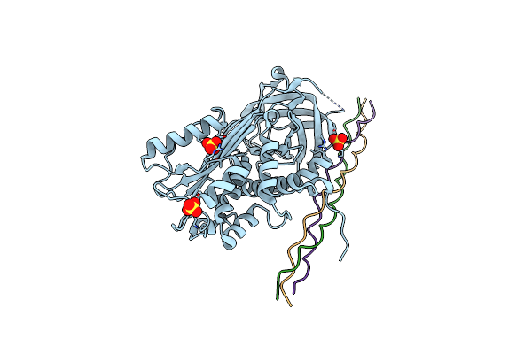
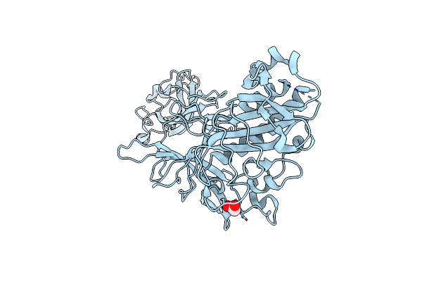
Crystal Structure Of Minor Pilin Cofb From Cfa/Iii Complexed With N-Terminal Peptide Fragment Of Cofj
Organism: Escherichia coli
Method: X-RAY DIFFRACTION Resolution:3.52 Å Release Date: 2018-06-27 Classification: CELL ADHESION |
 |
Organism: Escherichia coli
Method: X-RAY DIFFRACTION Resolution:1.76 Å Release Date: 2018-06-27 Classification: CELL ADHESION Ligands: CA |
 |
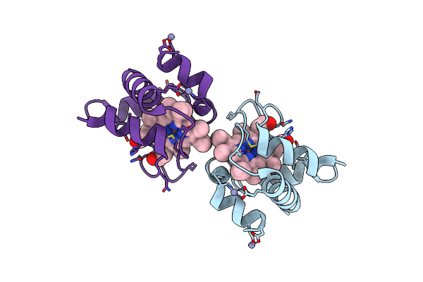
Homo-Dimeric Structure Of Cytochrome C' From Thermophilic Hydrogenophilus Thermoluteolus
Organism: Hydrogenophilus thermoluteolus
Method: X-RAY DIFFRACTION Resolution:1.89 Å Release Date: 2017-03-01 Classification: ELECTRON TRANSPORT Ligands: HEC |
 |
Organism: Clostridium perfringens
Method: X-RAY DIFFRACTION Resolution:1.89 Å Release Date: 2016-11-02 Classification: TOXIN |
 |
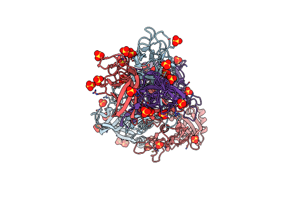
Crystal Structure Of An Adp-Ribosylating Toxin Beca Of A Novel Binary Enterotoxin Of C. Perfringens With Nadh
Organism: Clostridium perfringens
Method: X-RAY DIFFRACTION Resolution:1.83 Å Release Date: 2016-11-02 Classification: TOXIN Ligands: NAI |
 |
Organism: Shewanella violacea
Method: X-RAY DIFFRACTION Resolution:1.78 Å Release Date: 2016-10-19 Classification: ELECTRON TRANSPORT Ligands: HEC, IMD |
 |
The Crystal Structure Of Cofb, The Minor Pilin Subunit Of Cfa/Iii From Human Enterotoxigenic Escherichia Coli.
Organism: Escherichia coli
Method: X-RAY DIFFRACTION Resolution:1.88 Å Release Date: 2016-03-09 Classification: CELL ADHESION Ligands: ACT |