
Search Count: 37
 |
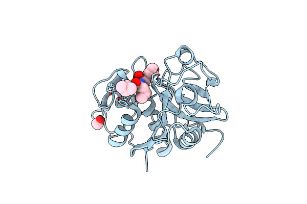
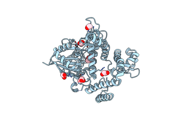
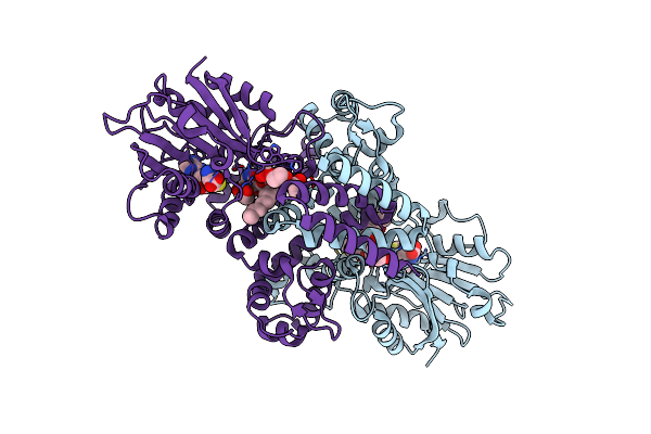
Organism: Sordaria araneosa
Method: X-RAY DIFFRACTION Release Date: 2025-10-22 Classification: BIOSYNTHETIC PROTEIN Ligands: A1LYT, MPD |
 |
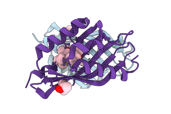
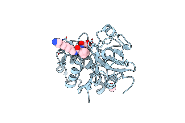
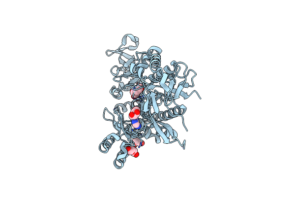
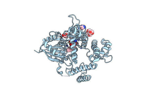
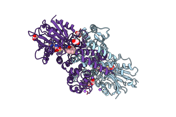
The Structure Of Sdng Covalently Binding With The Cope Rearrangement Product
Organism: Sordaria araneosa
Method: X-RAY DIFFRACTION Release Date: 2025-10-22 Classification: BIOSYNTHETIC PROTEIN Ligands: MPD, A1LZG |
 |
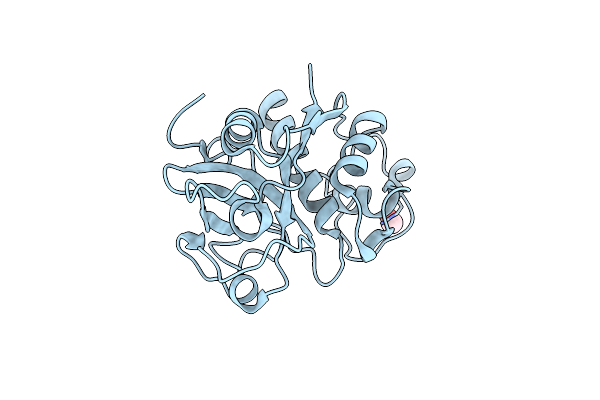
Organism: Sordaria araneosa
Method: X-RAY DIFFRACTION Release Date: 2025-10-22 Classification: BIOSYNTHETIC PROTEIN Ligands: A1LZH, A1LZK |
 |
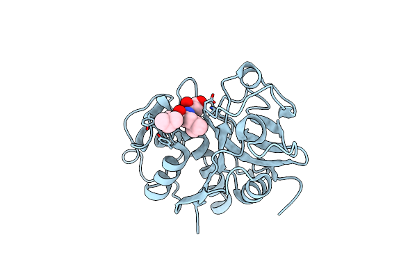
Organism: Sordaria araneosa
Method: X-RAY DIFFRACTION Release Date: 2025-10-22 Classification: BIOSYNTHETIC PROTEIN Ligands: MPD |
 |
Organism: Sordaria araneosa
Method: X-RAY DIFFRACTION Release Date: 2025-10-22 Classification: BIOSYNTHETIC PROTEIN Ligands: A1LYT |
 |
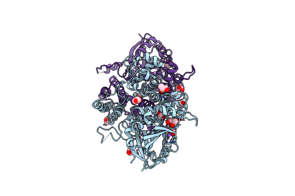
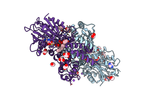
Organism: Carica papaya
Method: X-RAY DIFFRACTION Release Date: 2025-06-04 Classification: HYDROLASE Ligands: MOH, E64 |
 |
Organism: Carica papaya
Method: X-RAY DIFFRACTION Release Date: 2025-06-04 Classification: HYDROLASE Ligands: MOH, E6D |
 |
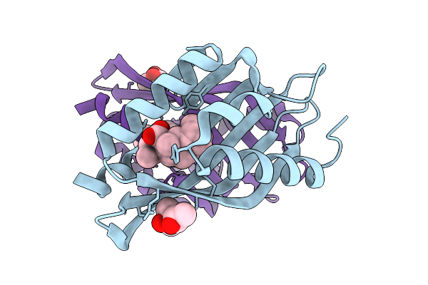
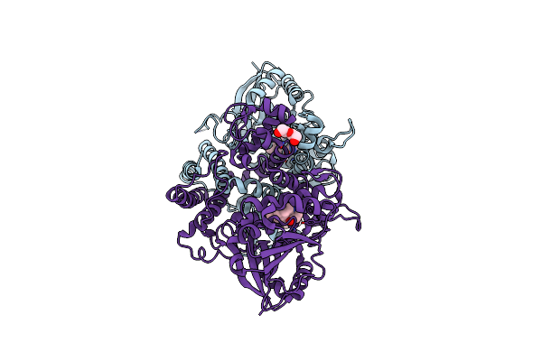
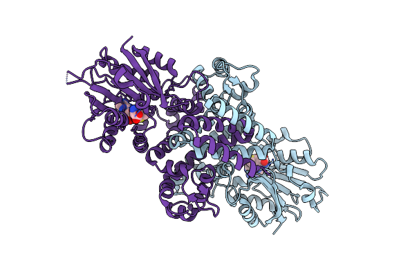
X-Ray Diffraction Structure Of Papain Co-Crystallized With Novel Biosynthetic Inhibitor Amine-65
Organism: Carica papaya
Method: X-RAY DIFFRACTION Release Date: 2025-06-04 Classification: HYDROLASE Ligands: MOH, A1AXI |
 |
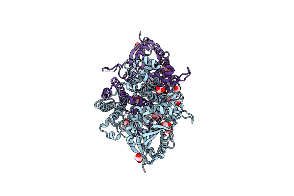
Organism: Carica papaya
Method: X-RAY DIFFRACTION Release Date: 2025-06-04 Classification: HYDROLASE Ligands: MOH |
 |
Organism: Carica papaya
Method: X-RAY DIFFRACTION Release Date: 2025-06-04 Classification: HYDROLASE Ligands: E6C |
 |
Organism: Aspergillus flavus
Method: X-RAY DIFFRACTION Release Date: 2025-05-21 Classification: LIGASE Ligands: MES, ADN, PEG |
 |
Organism: Aspergillus bombycis
Method: X-RAY DIFFRACTION Resolution:1.98 Å Release Date: 2020-10-14 Classification: BIOSYNTHETIC PROTEIN Ligands: EDO, GOL, NA |
 |
Organism: Aspergillus bombycis
Method: X-RAY DIFFRACTION Resolution:1.99 Å Release Date: 2020-10-14 Classification: BIOSYNTHETIC PROTEIN Ligands: F56, EDO, GOL |
 |
Organism: Aspergillus bombycis
Method: X-RAY DIFFRACTION Resolution:2.40 Å Release Date: 2020-10-14 Classification: BIOSYNTHETIC PROTEIN Ligands: F5F, PEG |
 |
Organism: Hymenoscyphus scutula
Method: X-RAY DIFFRACTION Resolution:1.53 Å Release Date: 2020-10-14 Classification: BIOSYNTHETIC PROTEIN Ligands: F56, IMD, GOL, B3P |
 |
Organism: Hymenoscyphus scutula
Method: X-RAY DIFFRACTION Resolution:1.33 Å Release Date: 2020-10-14 Classification: BIOSYNTHETIC PROTEIN Ligands: MPD, EDO, GOL |
 |
Organism: Aspergillus flavus
Method: X-RAY DIFFRACTION Resolution:2.13 Å Release Date: 2019-07-17 Classification: BIOSYNTHETIC PROTEIN Ligands: SAM, CL |
 |
Organism: Aspergillus flavus
Method: X-RAY DIFFRACTION Resolution:1.70 Å Release Date: 2019-07-17 Classification: BIOSYNTHETIC PROTEIN Ligands: SAM, B0L, EDO, CL, GOL, NA, IMD |
 |
Organism: Aspergillus flavus (strain atcc 200026 / fgsc a1120 / nrrl 3357 / jcm 12722 / srrc 167)
Method: X-RAY DIFFRACTION Resolution:1.84 Å Release Date: 2019-07-17 Classification: BIOSYNTHETIC PROTEIN Ligands: SAH, B0L, CL, GOL, EDO |
 |
Organism: Aspergillus flavus (strain atcc 200026 / fgsc a1120 / nrrl 3357 / jcm 12722 / srrc 167)
Method: X-RAY DIFFRACTION Resolution:1.66 Å Release Date: 2019-07-17 Classification: BIOSYNTHETIC PROTEIN Ligands: SAM, CL, NA, B3O, EDO, ACT |