
Search Count: 49
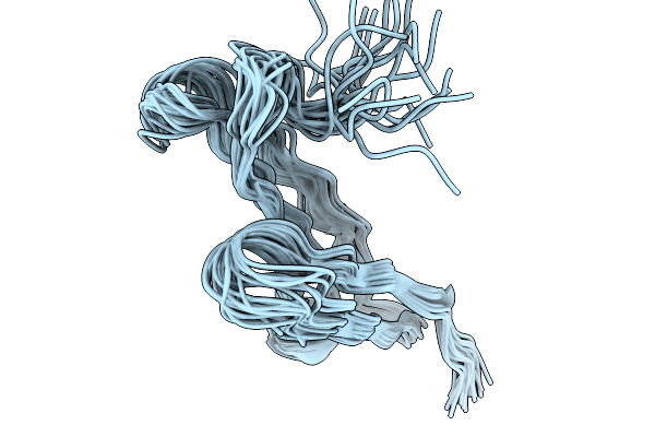 |
Organism: Colletotrichum orbiculare
Method: SOLUTION NMR Release Date: 2025-11-19 Classification: VIRAL PROTEIN |
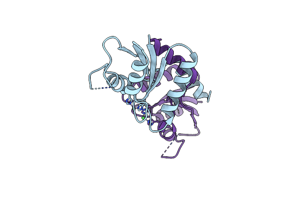 |
Organism: Methanocaldococcus jannaschii (strain atcc 43067 / dsm 2661 / jal-1 / jcm 10045 / nbrc 100440)
Method: X-RAY DIFFRACTION Resolution:2.10 Å Release Date: 2024-04-10 Classification: BIOSYNTHETIC PROTEIN Ligands: NI |
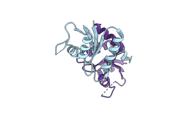 |
Organism: Methanocaldococcus jannaschii dsm 2661
Method: X-RAY DIFFRACTION Resolution:1.99 Å Release Date: 2024-01-31 Classification: BIOSYNTHETIC PROTEIN |
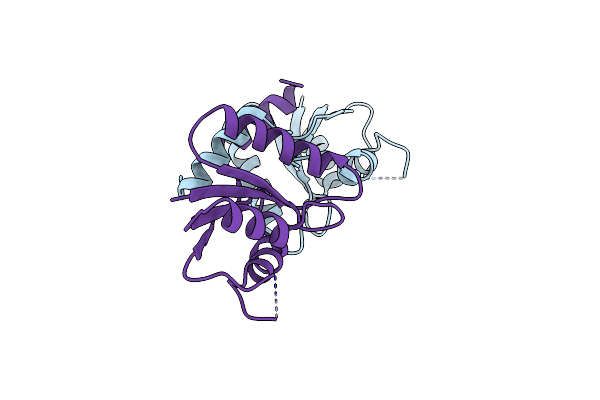 |
Organism: Methanocaldococcus jannaschii (strain atcc 43067 / dsm 2661 / jal-1 / jcm 10045 / nbrc 100440)
Method: X-RAY DIFFRACTION Resolution:2.84 Å Release Date: 2024-01-31 Classification: BIOSYNTHETIC PROTEIN |
 |
Organism: Methanocaldococcus jannaschii (strain atcc 43067 / dsm 2661 / jal-1 / jcm 10045 / nbrc 100440)
Method: X-RAY DIFFRACTION Resolution:2.81 Å Release Date: 2024-01-31 Classification: BIOSYNTHETIC PROTEIN Ligands: UP3 |
 |
Organism: Methanocaldococcus jannaschii (strain atcc 43067 / dsm 2661 / jal-1 / jcm 10045 / nbrc 100440)
Method: X-RAY DIFFRACTION Resolution:3.09 Å Release Date: 2024-01-31 Classification: BIOSYNTHETIC PROTEIN Ligands: UPI |
 |
Crystal Structure Of Cysteine Desulfurase Sufs C361A From Bacillus Subtilis
Organism: Bacillus subtilis subsp. subtilis str. 168
Method: X-RAY DIFFRACTION Resolution:1.96 Å Release Date: 2022-03-02 Classification: BIOSYNTHETIC PROTEIN Ligands: EDO, PEG, PG4 |
 |
Crystal Structure Of Pmp-Bound Form Of Cysteine Desulfurase Sufs C361A From Bacillus Subtilis
Organism: Bacillus subtilis subsp. subtilis str. 168
Method: X-RAY DIFFRACTION Resolution:1.84 Å Release Date: 2022-03-02 Classification: BIOSYNTHETIC PROTEIN Ligands: PMP, EDO, PEG, PG4 |
 |
Crystal Structure Of L-Cycloserine-Bound Form Of Cysteine Desulfurase Sufs C361A From Bacillus Subtilis
Organism: Bacillus subtilis subsp. subtilis str. 168
Method: X-RAY DIFFRACTION Resolution:1.73 Å Release Date: 2022-03-02 Classification: BIOSYNTHETIC PROTEIN Ligands: PEG, EDO, PG4, 7TS |
 |
Crystal Structure Of Cysteine Desulfurase Sufs R376A From Bacillus Subtilis
Organism: Bacillus subtilis subsp. subtilis str. 168
Method: X-RAY DIFFRACTION Resolution:2.67 Å Release Date: 2022-03-02 Classification: BIOSYNTHETIC PROTEIN Ligands: PEG |
 |
Crystal Structure Of Pmp-Bound Form Of Cysteine Desulfurase Sufs R376A From Bacillus Subtilis In D-Cycloserine-Inhibition
Organism: Bacillus subtilis subsp. subtilis str. 168
Method: X-RAY DIFFRACTION Resolution:2.28 Å Release Date: 2022-03-02 Classification: BIOSYNTHETIC PROTEIN Ligands: PMP, PG4, PEG, EDO |
 |
Crystal Structure Of Pmp-Bound Form Of Cysteine Desulfurase Sufs R376A From Bacillus Subtilis In L-Cycloserine-Inhibition
Organism: Bacillus subtilis subsp. subtilis str. 168
Method: X-RAY DIFFRACTION Resolution:2.74 Å Release Date: 2022-03-02 Classification: BIOSYNTHETIC PROTEIN Ligands: PEG, PMP, EDO |
 |
Organism: Colletotrichum orbiculare (strain 104-t / atcc 96160 / cbs 514.97 / lars 414 / maff 240422)
Method: SOLUTION NMR Release Date: 2021-11-03 Classification: TOXIN |
 |
Crystal Structure Of Pmp-Bound Form Of Cysteine Desulfurase Sufs From Bacillus Subtilis
Organism: Bacillus subtilis subsp. subtilis str. 168
Method: X-RAY DIFFRACTION Resolution:2.43 Å Release Date: 2021-06-30 Classification: BIOSYNTHETIC PROTEIN Ligands: PEG, PMP |
 |
Crystal Structure Of L-Cycloserine-Bound Form Of Cysteine Desulfurase Sufs From Bacillus Subtilis
Organism: Bacillus subtilis subsp. subtilis str. 168
Method: X-RAY DIFFRACTION Resolution:2.05 Å Release Date: 2021-06-30 Classification: BIOSYNTHETIC PROTEIN Ligands: PEG, 7TS, PGE |
 |
Crystal Structure Of Cysteine Desulfurase Sufs H121A From Bacillus Subtilis
Organism: Bacillus subtilis subsp. subtilis str. 168
Method: X-RAY DIFFRACTION Resolution:2.00 Å Release Date: 2021-06-30 Classification: BIOSYNTHETIC PROTEIN Ligands: PEG |
 |
Crystal Structure Of D-Cycloserine-Bound Form Of Cysteine Desulfurase Sufs H121A From Bacillus Subtilis
Organism: Bacillus subtilis subsp. subtilis str. 168
Method: X-RAY DIFFRACTION Resolution:2.30 Å Release Date: 2021-06-30 Classification: BIOSYNTHETIC PROTEIN Ligands: DCS, PGE, PEG |
 |
Crystal Structure Of L-Cycloserine-Bound Form Of Cysteine Desulfurase Sufs H121A From Bacillus Subtilis
Organism: Bacillus subtilis subsp. subtilis str. 168
Method: X-RAY DIFFRACTION Resolution:2.20 Å Release Date: 2021-06-30 Classification: BIOSYNTHETIC PROTEIN Ligands: PEG, 7TS |
 |
Crystal Structure Of D-Cycloserine-Bound Form Of Cysteine Desulfurase Nifs From Helicobacter Pylori
Organism: Helicobacter pylori 26695
Method: X-RAY DIFFRACTION Resolution:2.64 Å Release Date: 2021-06-30 Classification: BIOSYNTHETIC PROTEIN Ligands: IPA, CL, 7TS |
 |
Crystal Structure Of L-Cycloserine-Bound Form Of Cysteine Desulfurase Nifs From Helicobacter Pylori
Organism: Helicobacter pylori 26695
Method: X-RAY DIFFRACTION Resolution:2.90 Å Release Date: 2021-06-30 Classification: BIOSYNTHETIC PROTEIN Ligands: IPA, 7TS |