
Search Count: 113
 |
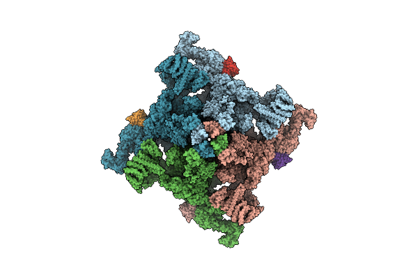
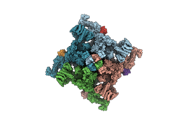
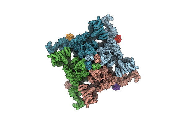
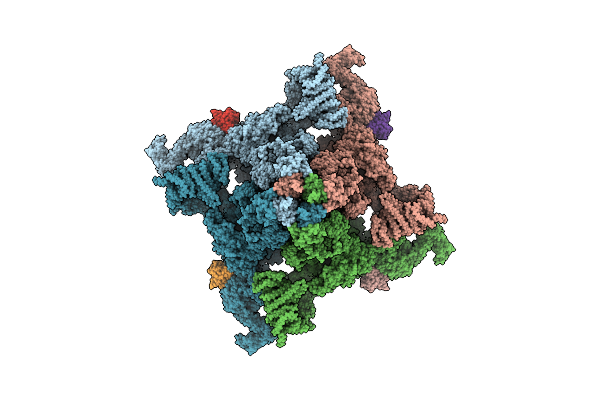
Organism: Thalassiosira pseudonana ccmp1335
Method: ELECTRON MICROSCOPY Release Date: 2024-10-30 Classification: PHOTOSYNTHESIS Ligands: CL0, CLA, PQN, SF4, BCR, LHG, LMT, UNL, DGD, 5X6, DD6, LMG, KC1, A86 |
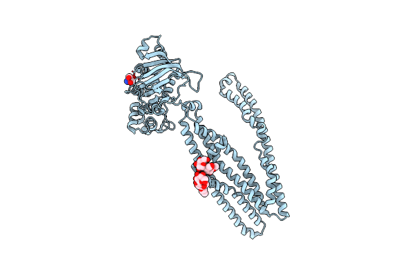 |
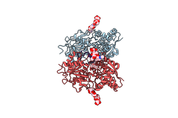
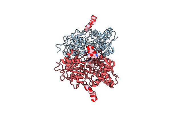
Crystal Structure Of Cmabcb1 W114Y/W161Y/W363Y/W364Y/M391W (4Wy/M391W) Mutant
Organism: Cyanidioschyzon merolae (strain 10d)
Method: X-RAY DIFFRACTION Resolution:3.00 Å Release Date: 2022-09-07 Classification: TRANSPORT PROTEIN Ligands: DMU, TRS |
 |
Organism: Mus musculus, Homo sapiens
Method: ELECTRON MICROSCOPY Release Date: 2022-08-10 Classification: MEMBRANE PROTEIN Ligands: ZN |
 |
Organism: Mus musculus, Homo sapiens
Method: ELECTRON MICROSCOPY Release Date: 2022-08-10 Classification: MEMBRANE PROTEIN Ligands: ZN |
 |
Organism: Mus musculus, Homo sapiens
Method: ELECTRON MICROSCOPY Release Date: 2022-08-10 Classification: MEMBRANE PROTEIN Ligands: ZN |
 |
Organism: Mus musculus, Homo sapiens
Method: ELECTRON MICROSCOPY Release Date: 2022-08-10 Classification: MEMBRANE PROTEIN Ligands: ZN, CA |
 |
Organism: Mus musculus, Homo sapiens
Method: ELECTRON MICROSCOPY Release Date: 2022-08-10 Classification: MEMBRANE PROTEIN Ligands: ZN, CA |
 |
Organism: Mus musculus, Homo sapiens
Method: ELECTRON MICROSCOPY Release Date: 2022-08-10 Classification: MEMBRANE PROTEIN Ligands: ZN, CA |
 |
Organism: Mus musculus, Homo sapiens
Method: ELECTRON MICROSCOPY Release Date: 2022-08-10 Classification: MEMBRANE PROTEIN Ligands: ZN |
 |
Organism: Mus musculus, Homo sapiens
Method: ELECTRON MICROSCOPY Release Date: 2022-08-10 Classification: MEMBRANE PROTEIN Ligands: ZN, CA |
 |
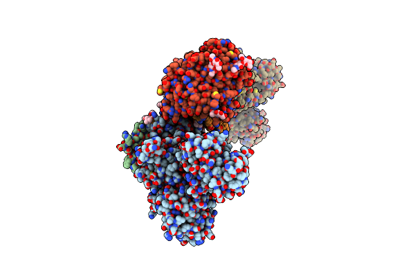
Atrial Natriuretic Peptide Receptor Complexed With Rat Atrial Natriuretic Peptide
Organism: Rattus norvegicus
Method: X-RAY DIFFRACTION Resolution:2.45 Å Release Date: 2021-03-31 Classification: MEMBRANE PROTEIN Ligands: CL |
 |
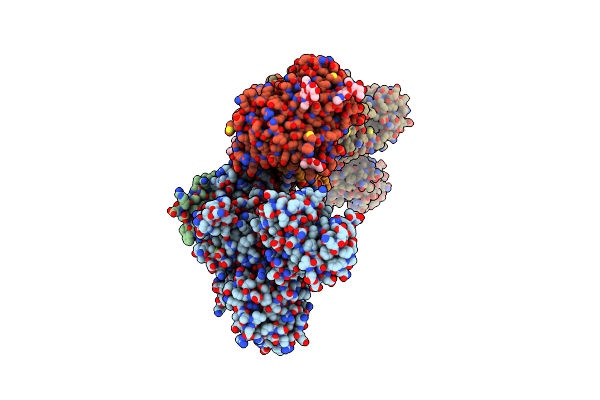
Atrial Natriuretic Peptide Receptor Complexed With Human Atrial Natriuretic Peptide
Organism: Rattus norvegicus, Homo sapiens
Method: X-RAY DIFFRACTION Resolution:2.45 Å Release Date: 2021-03-31 Classification: MEMBRANE PROTEIN Ligands: CL |
 |
Atrial Natriuretic Peptide Receptor Complexed With Dendroaspis Natriuretic Peptide
Organism: Rattus norvegicus, Dendroaspis angusticeps
Method: X-RAY DIFFRACTION Resolution:2.45 Å Release Date: 2021-03-31 Classification: MEMBRANE PROTEIN Ligands: CL |
 |
Atrial Natriuretic Peptide Receptor Complexed With Deletion Mutant Of Human Atrial Natriuretic Peptide[7-28]
Organism: Rattus norvegicus, Homo sapiens
Method: X-RAY DIFFRACTION Resolution:2.70 Å Release Date: 2021-03-31 Classification: MEMBRANE PROTEIN Ligands: CL |
 |
Atrial Natriuretic Peptide Receptor Complexed With Deletion Mutant Of Human Atrial Natriuretic Peptide[5-27]
Organism: Rattus norvegicus, Homo sapiens
Method: X-RAY DIFFRACTION Resolution:2.85 Å Release Date: 2021-03-31 Classification: MEMBRANE PROTEIN Ligands: CL |
 |
Atrial Natriuretic Peptide Receptor Complexed With Deletion Mutant Of Rat Atrial Natriuretic Peptide[4-17,23]
Organism: Rattus norvegicus
Method: X-RAY DIFFRACTION Resolution:3.20 Å Release Date: 2021-03-31 Classification: MEMBRANE PROTEIN Ligands: CL |
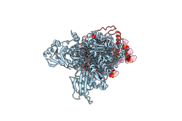 |
Crystal Structure Of The Na+,K+-Atpase In The E2P State With Bound Mg2+ (P4(3)2(1)2 Symmetry)
Organism: Sus scrofa
Method: X-RAY DIFFRACTION Resolution:3.35 Å Release Date: 2021-01-27 Classification: TRANSPORT PROTEIN Ligands: MG, CLR, PCW, NAG |
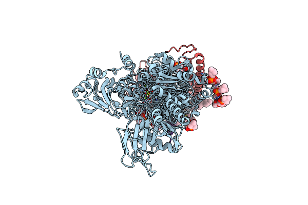 |
Crystal Structure Of The Na+,K+-Atpase In The E2P State With Bound Mg2+ And Anthroylouabain (P4(3)2(1)2 Symmetry)
Organism: Sus scrofa
Method: X-RAY DIFFRACTION Resolution:3.90 Å Release Date: 2021-01-27 Classification: TRANSPORT PROTEIN Ligands: MG, CLR, PCW, H0C, NAG |
 |
Crystal Structure Of The Na+,K+-Atpase In The E2P State With Bound Mg2+ And Anthroylouabain (P2(1)2(1)2(1) Symmetry)
Organism: Sus scrofa
Method: X-RAY DIFFRACTION Resolution:3.65 Å Release Date: 2021-01-27 Classification: TRANSPORT PROTEIN Ligands: MG, NA, CLR, PCW, H0C, NAG |
 |
Crystal Structure Of The Na+,K+-Atpase In The E2P State With Bound One Mg2+ And One Rb+ In The Presence Of Bufalin
Organism: Sus scrofa
Method: X-RAY DIFFRACTION Resolution:3.50 Å Release Date: 2021-01-27 Classification: TRANSPORT PROTEIN Ligands: MG, NA, RB, CLR, PCW, BUF, NAG |