
Search Count: 44
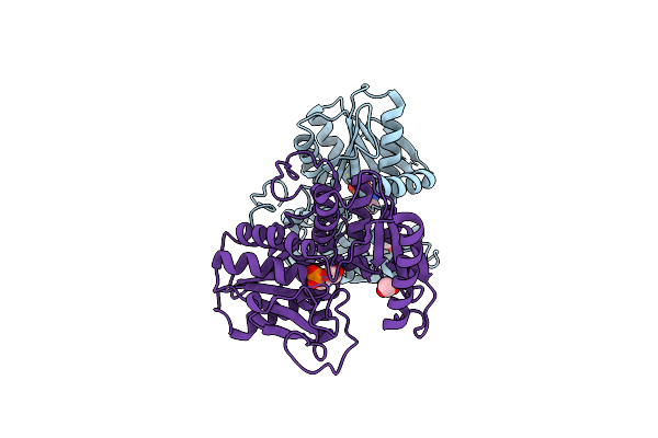 |
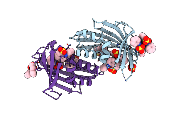
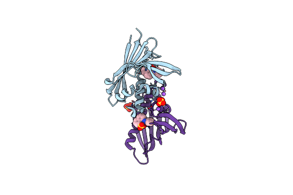
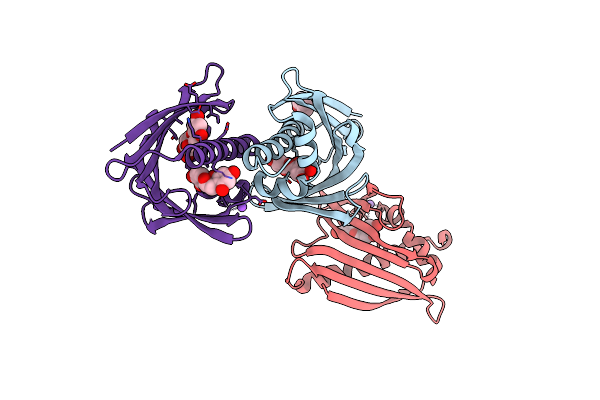
Structural Characterization Of Beta Cyanoalanine Synthase From Tetranychus Urticae
Organism: Tetranychus urticae
Method: X-RAY DIFFRACTION Resolution:2.35 Å Release Date: 2021-11-24 Classification: STRUCTURAL PROTEIN Ligands: PLP, ACT |
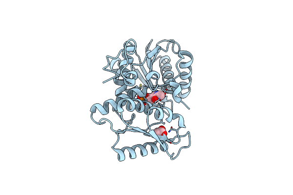 |
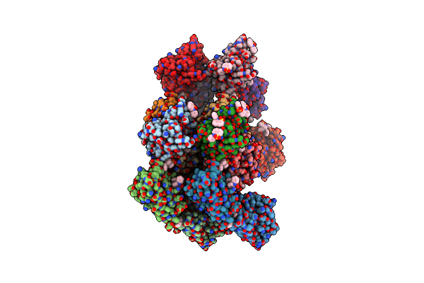
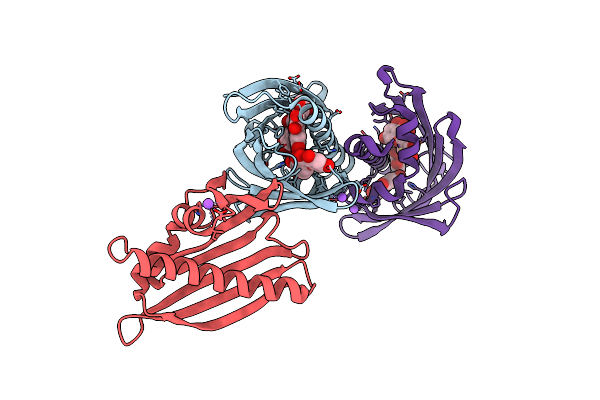
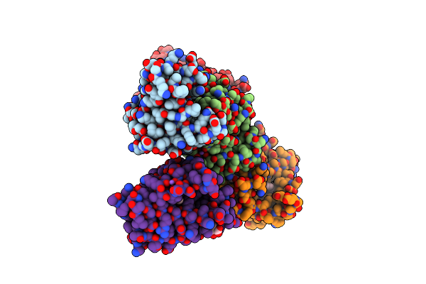
Structural Characterization Of Beta Cyanoalanine Synthase From Tetranychus Urticae (Two-Spotted Spider Mite)
Organism: Tetranychus urticae
Method: X-RAY DIFFRACTION Resolution:1.60 Å Release Date: 2021-05-19 Classification: BIOSYNTHETIC PROTEIN Ligands: PLP, ACT |
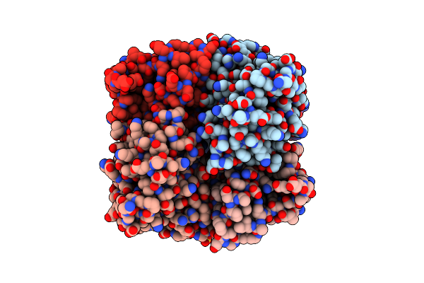 |
Organism: Arachis hypogaea
Method: X-RAY DIFFRACTION Resolution:2.31 Å Release Date: 2020-12-16 Classification: ALLERGEN Ligands: SO4 |
 |
Organism: Arachis hypogaea
Method: X-RAY DIFFRACTION Resolution:1.95 Å Release Date: 2020-12-16 Classification: ALLERGEN |
 |
Organism: Arachis hypogaea
Method: X-RAY DIFFRACTION Resolution:1.95 Å Release Date: 2020-12-16 Classification: ALLERGEN Ligands: SO4, DCR |
 |
Organism: Arachis hypogaea
Method: X-RAY DIFFRACTION Resolution:1.75 Å Release Date: 2020-12-16 Classification: ALLERGEN Ligands: 2AN, SO4 |
 |
Organism: Arachis hypogaea
Method: X-RAY DIFFRACTION Resolution:2.10 Å Release Date: 2020-12-16 Classification: ALLERGEN Ligands: 2AN, SO4 |
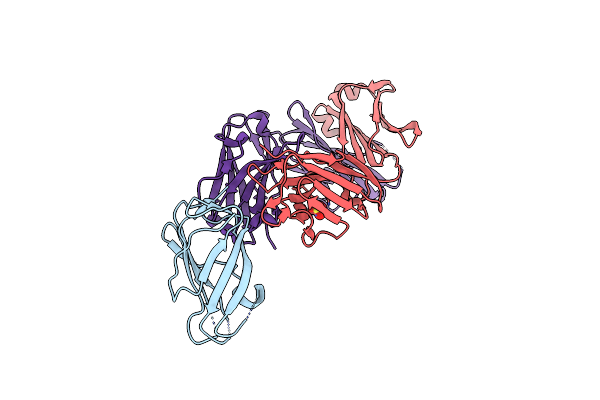 |
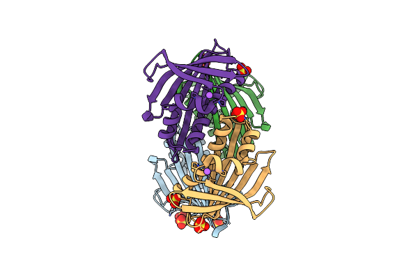
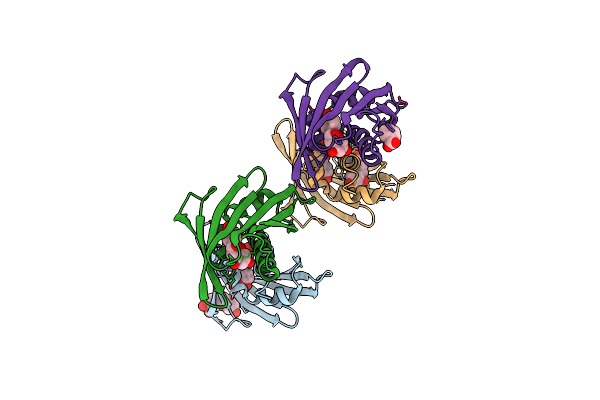
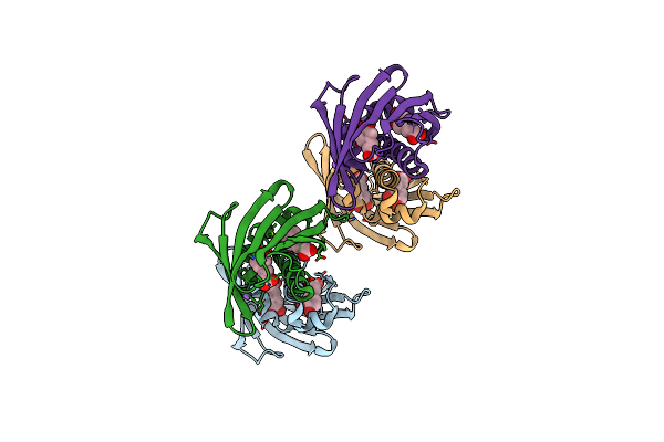
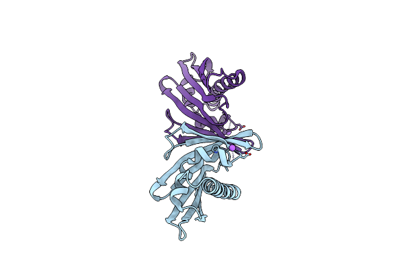
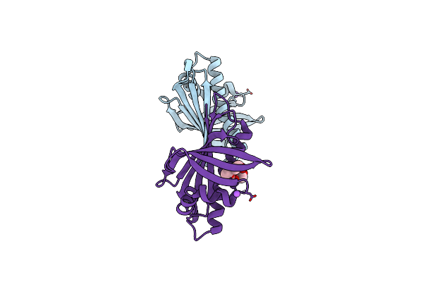
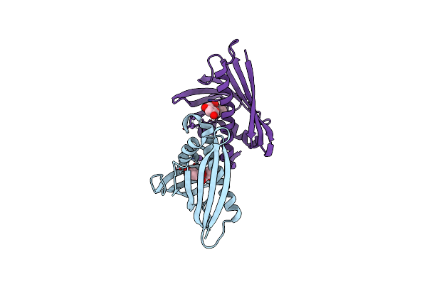
Crystal Structure Of Complex Between Recombinant Der P 2.0103 And Fab Fragment Of 7A1
Organism: Dermatophagoides pteronyssinus, Mus musculus
Method: X-RAY DIFFRACTION Resolution:2.45 Å Release Date: 2019-08-28 Classification: allergen/immune system Ligands: SO4 |
 |
Organism: Arachis hypogaea
Method: X-RAY DIFFRACTION Resolution:2.51 Å Release Date: 2018-09-19 Classification: PROTEIN BINDING Ligands: QUE, BGC |
 |
Organism: Arachis hypogaea
Method: X-RAY DIFFRACTION Resolution:1.60 Å Release Date: 2018-09-12 Classification: PROTEIN BINDING Ligands: 2AN, NA, SO4 |
 |
Organism: Arachis hypogaea
Method: X-RAY DIFFRACTION Resolution:2.35 Å Release Date: 2018-09-12 Classification: PROTEIN BINDING |
 |
Organism: Arachis hypogaea
Method: X-RAY DIFFRACTION Resolution:2.30 Å Release Date: 2018-09-12 Classification: PLANT PROTEIN Ligands: 28E, NA, BEZ |
 |
Organism: Arachis hypogaea
Method: X-RAY DIFFRACTION Resolution:3.05 Å Release Date: 2018-09-12 Classification: PROTEIN BINDING Ligands: DHC, NA, CL |
 |
Organism: Arachis hypogaea
Method: X-RAY DIFFRACTION Resolution:2.55 Å Release Date: 2018-09-12 Classification: PROTEIN BINDING Ligands: 28E, BEZ, NA |
 |
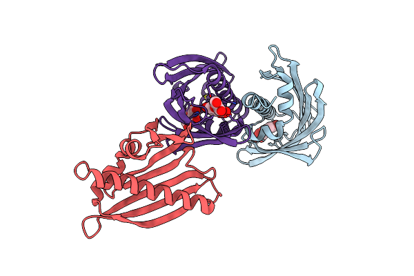
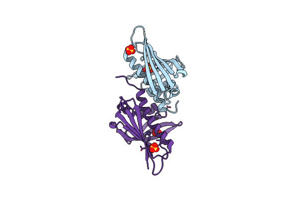
Structure Of Pr 10 Allergen Ara H 8.01 In Complex With 3-Hydroxy-2-Naphthoic Acid
Organism: Arachis hypogaea
Method: X-RAY DIFFRACTION Resolution:2.30 Å Release Date: 2018-09-12 Classification: PROTEIN BINDING |
 |
Organism: Arachis hypogaea
Method: X-RAY DIFFRACTION Resolution:2.70 Å Release Date: 2018-09-12 Classification: PLANT PROTEIN |
 |
Organism: Arachis hypogaea
Method: X-RAY DIFFRACTION Resolution:2.10 Å Release Date: 2018-09-12 Classification: PROTEIN BINDING Ligands: SO4, NA |
 |
Organism: Arachis hypogaea
Method: X-RAY DIFFRACTION Resolution:1.90 Å Release Date: 2018-09-12 Classification: PLANT PROTEIN |
 |
Organism: Arachis hypogaea
Method: X-RAY DIFFRACTION Resolution:2.50 Å Release Date: 2018-09-12 Classification: PROTEIN BINDING |
 |
Organism: Vibrio vulnificus (strain cmcp6)
Method: X-RAY DIFFRACTION Resolution:2.35 Å Release Date: 2017-07-12 Classification: OXIDOREDUCTASE Ligands: PO4, EDO |