
Search Count: 7
All
Selected
 |
Organism: Homo sapiens
Method: X-RAY DIFFRACTION Resolution:2.50 Å Release Date: 2022-03-30 Classification: OXIDOREDUCTASE |
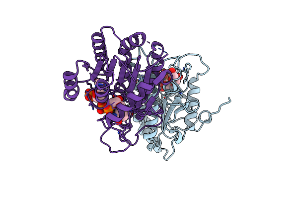 |
Crystal Structure Of Nadph Bound Carbonyl Reductase From Chicken Fatty Liver
Organism: Gallus gallus
Method: X-RAY DIFFRACTION Resolution:1.98 Å Release Date: 2015-07-29 Classification: OXIDOREDUCTASE ACTIVATOR Ligands: NDP, EDO |
 |
Organism: Gluconobacter oxydans
Method: X-RAY DIFFRACTION Resolution:3.30 Å Release Date: 2014-09-24 Classification: OXIDOREDUCTASE ACTIVATOR Ligands: PGE, PEG, HG |
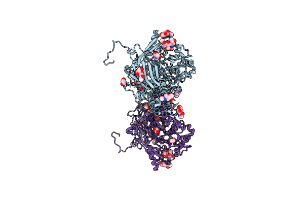 |
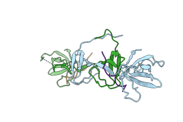
Crystal Structure Of Hemolymph Type Prophenoloxidase (Propob) From Crustacean
Organism: Marsupenaeus japonicus
Method: X-RAY DIFFRACTION Resolution:1.80 Å Release Date: 2014-04-23 Classification: OXIDOREDUCTASE ACTIVATOR Ligands: NAG, CUO, EDO, NA, CL, MG |
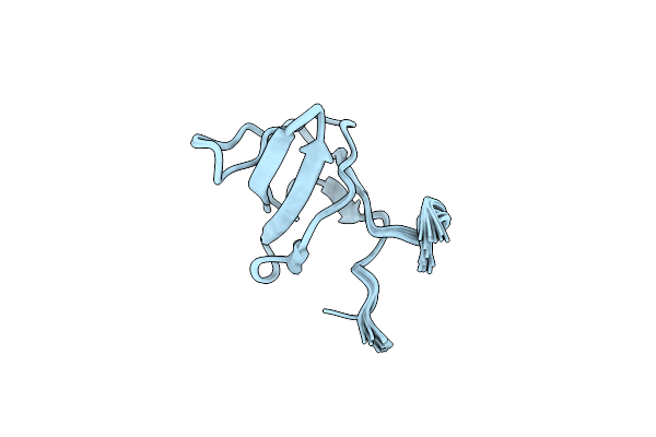 |
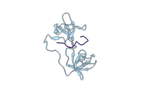
Organism: Homo sapiens
Method: SOLUTION NMR Release Date: 2006-03-21 Classification: Oxidoreductase Activator |
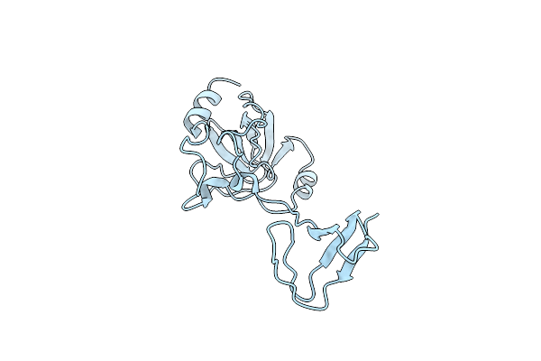 |
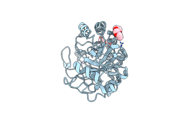
Organism: Homo sapiens
Method: X-RAY DIFFRACTION Resolution:1.70 Å Release Date: 2003-05-20 Classification: Oxidoreductase activator |
 |
Organism: Homo sapiens
Method: X-RAY DIFFRACTION Resolution:1.80 Å Release Date: 2003-05-20 Classification: Oxidoreductase activator |