
Search Count: 19
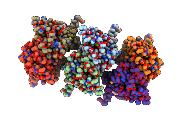 |
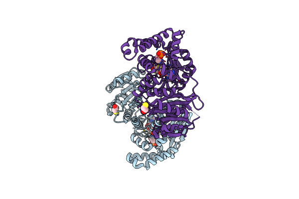
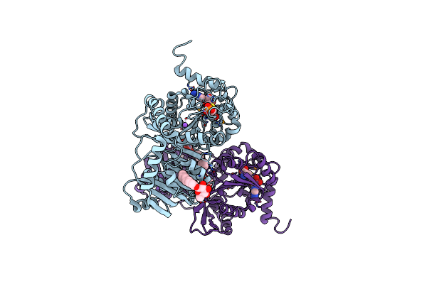
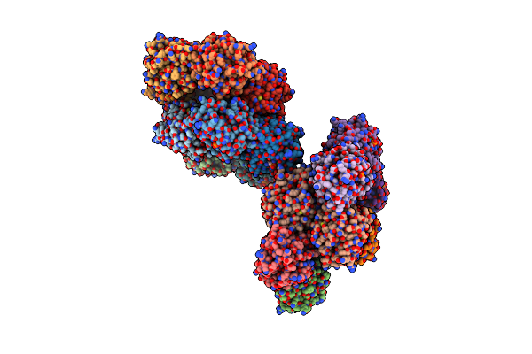
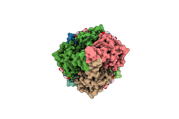
The Cryo-Em Structure Of Human Sphingomyelin Synthase-Related Protein In Complex With Diacylglycerol/Phosphoethanolamine
Organism: Homo sapiens
Method: ELECTRON MICROSCOPY Release Date: 2024-02-28 Classification: MEMBRANE PROTEIN Ligands: Z0P, OPE |
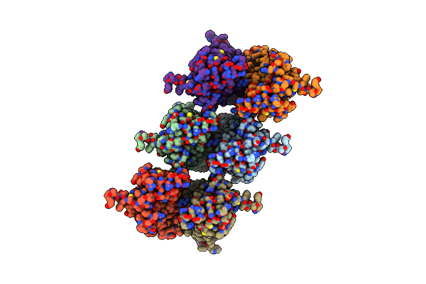 |
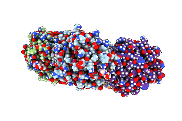
The Cryo-Em Structure Of Human Sphingomyelin Synthase-Related Protein In Complex With Ceramide/Phosphoethanolamine
Organism: Homo sapiens
Method: ELECTRON MICROSCOPY Release Date: 2024-02-28 Classification: MEMBRANE PROTEIN Ligands: UJO, OPE |
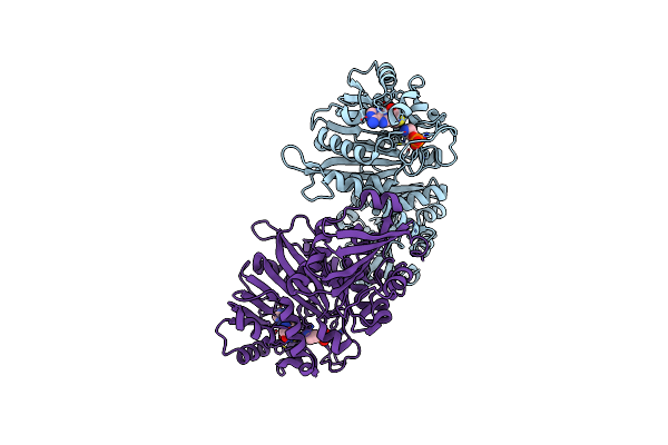 |
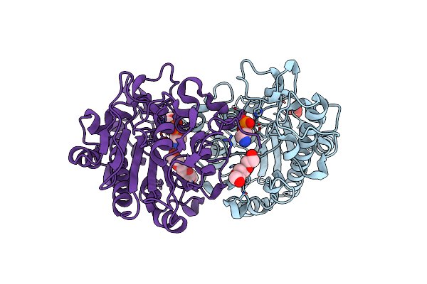
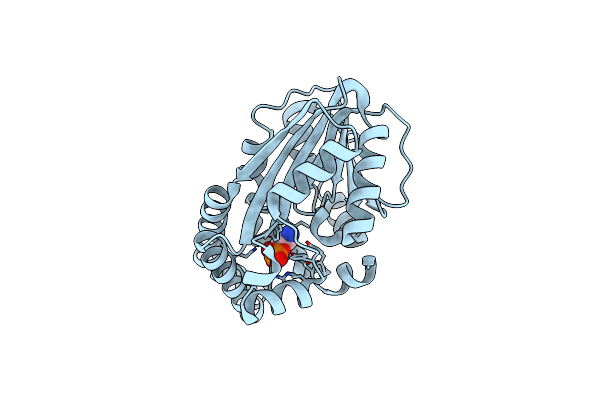
Phosphoethanolamine Methyltransferase From The Pine Wilt Nematode Bursaphelenchus Xylophilus
Organism: Bursaphelenchus xylophilus
Method: X-RAY DIFFRACTION Resolution:2.05 Å Release Date: 2020-06-17 Classification: TRANSFERASE Ligands: OPE, SAH |
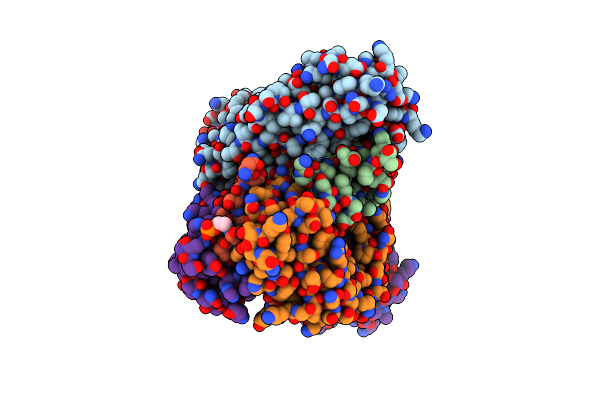 |
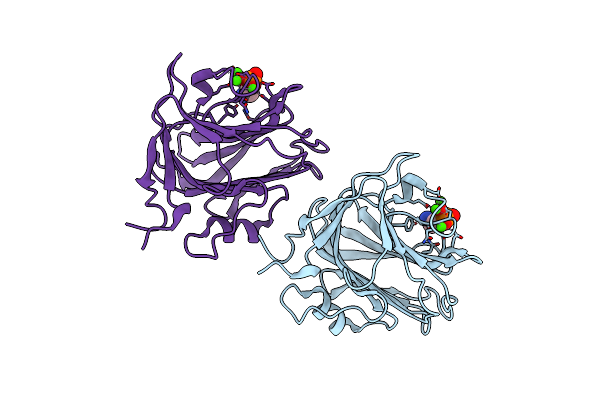
Crystal Structure Of Anti-Gld52 Fab Complex With Human Gld52 Peptide Mimetic
Organism: Mus musculus, Homo sapiens
Method: X-RAY DIFFRACTION Resolution:2.20 Å Release Date: 2019-07-03 Classification: IMMUNE SYSTEM Ligands: ZN, OPE |
 |
Crystal Structure Of The Intrinsic Colistin Resistance Enzyme Icr(Mc) From Moraxella Catarrhalis, Catalytic Domain, Thr315Ala Mutant Mono-Zinc And Phosphoethanolamine Complex
Organism: Moraxella sp. hmsc061h09
Method: X-RAY DIFFRACTION Resolution:1.66 Å Release Date: 2018-01-31 Classification: TRANSFERASE Ligands: ZN, OPE, SO4, 15P |
 |
Organism: Neisseria gonorrhoeae
Method: ELECTRON MICROSCOPY Resolution:5.10 Å Release Date: 2017-07-12 Classification: PROTEIN FIBRIL Ligands: OPE |
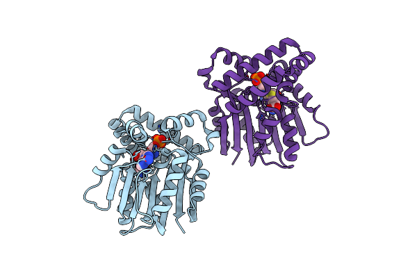 |
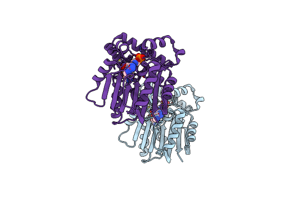
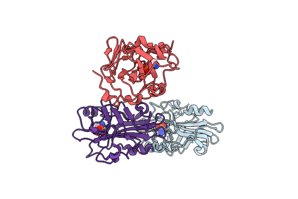
Plasmodium Falciparum Phosphoethanolamine Methyltransferase D128A Mutant In Complex With S-Adenosylhomocysteine And Phosphoethanolamine
Organism: Plasmodium falciparum
Method: X-RAY DIFFRACTION Resolution:2.55 Å Release Date: 2014-10-08 Classification: TRANSFERASE Ligands: OPE, SAH |
 |
Crystal Structure Of N-Methyl Transferase (Pmt-2) From Caenorhabditis Elegant Complexed With S-Adenosyl Homocysteine And Phosphoethanolamine
Organism: Caenorhabditis elegans
Method: X-RAY DIFFRACTION Resolution:1.45 Å Release Date: 2014-01-15 Classification: TRANSFERASE Ligands: SAH, OPE, BME |
 |
Semet Haemonchus Contortus Phosphoethanolamine N-Methyltransferase 1 In Complex With Phosphoethanolamine And S-Adenosylhomocysteine
Organism: Haemonchus contortus
Method: X-RAY DIFFRACTION Resolution:1.68 Å Release Date: 2013-09-25 Classification: TRANSFERASE Ligands: OPE, SAH, BOG, NA |
 |
Phosphoethanolamine Methyltransferase From Plasmodium Falciparum In Complex With Phosphoethanolamine
Organism: Plasmodium falciparum
Method: X-RAY DIFFRACTION Resolution:1.47 Å Release Date: 2011-11-30 Classification: TRANSFERASE Ligands: OPE |
 |
Phosphoethanolamine Methyltransferase From Plasmodium Falciparum In Complex With Sah And Phosphoethanolamine
Organism: Plasmodium falciparum
Method: X-RAY DIFFRACTION Resolution:1.52 Å Release Date: 2011-11-30 Classification: TRANSFERASE Ligands: OPE, SAH |
 |
Organism: Homo sapiens
Method: X-RAY DIFFRACTION Resolution:1.50 Å Release Date: 2010-12-08 Classification: GLYCOPROTEIN Ligands: OPE, CA, NAG |
 |
Organism: Homo sapiens
Method: X-RAY DIFFRACTION Resolution:2.70 Å Release Date: 2010-12-08 Classification: Calcium-BINDING PROTEIN Ligands: CA, OPE |
 |
Organism: Plasmodium falciparum 3d7
Method: X-RAY DIFFRACTION Resolution:2.30 Å Release Date: 2009-02-10 Classification: TRANSFERASE Ligands: ADP, MG, OPE |
 |
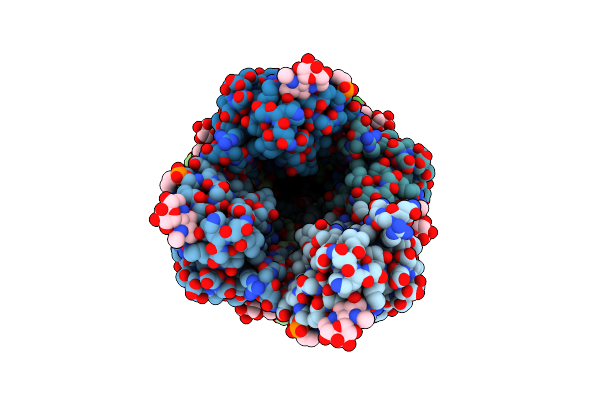
Crystal Structure Of Pe-Bound Octameric Sap-Like Pentraxin From Limulus Polyphemus
Organism: Limulus polyphemus
Method: X-RAY DIFFRACTION Resolution:2.70 Å Release Date: 2009-01-20 Classification: SUGAR BINDING PROTEIN Ligands: CA, OPE |
 |
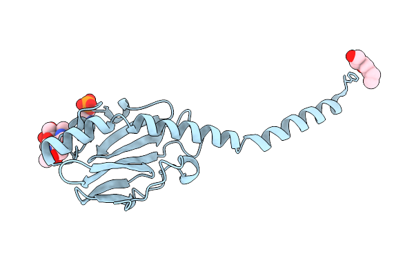
Rat Phosphatidylethanolamine-Binding Protein Containing The S153E Mutation In The Complex With O-Phosphorylethanolamine
Organism: Rattus norvegicus
Method: X-RAY DIFFRACTION Resolution:2.20 Å Release Date: 2007-09-25 Classification: HYDROLASE INHIBITOR Ligands: OPE |
 |
Crystal Structure Of Native Neisseria Gonorrhoeae Type Iv Pilin At 2.3 Angstroms Resolution
Organism: Neisseria gonorrhoeae
Method: X-RAY DIFFRACTION Resolution:2.30 Å Release Date: 2006-09-12 Classification: CELL ADHESION Ligands: HTO, OPE |
 |
Structure Of The Neisseria Gonorrhoeae Type Iv Pilus Filament From X-Ray Crystallography And Electron Cryomicroscopy
Organism: Neisseria gonorrhoeae
Method: ELECTRON MICROSCOPY Resolution:12.50 Å Release Date: 2006-09-12 Classification: CELL ADHESION Ligands: OPE |
 |
Structure Of The Phosphatidylethanolamine-Binding Protein From Bovine Brain
Organism: Bos taurus
Method: X-RAY DIFFRACTION Resolution:2.25 Å Release Date: 1999-01-27 Classification: LIPID BINDING PROTEIN Ligands: OPE |