
Search Count: 23
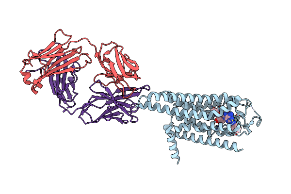 |
Crystal Structure Of The Human Adenosine A2A Receptor In Complex With Photoresponsive Ligand Photoneca(Blue)
Organism: Homo sapiens, Mus musculus
Method: X-RAY DIFFRACTION Resolution:3.34 Å Release Date: 2024-01-17 Classification: MEMBRANE PROTEIN Ligands: WCH |
 |
Organism: Aequorea victoria
Method: X-RAY DIFFRACTION Resolution:2.02 Å Release Date: 2023-08-02 Classification: FLUORESCENT PROTEIN |
 |
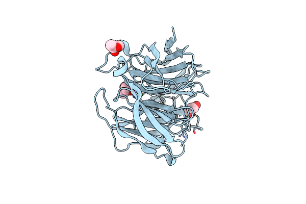
Room Temperature Rsegfp2 With A Chlorinated Chromophore In The Non-Fluorescent Off-State
Organism: Aequorea victoria
Method: X-RAY DIFFRACTION Resolution:1.63 Å Release Date: 2023-07-19 Classification: FLUORESCENT PROTEIN |
 |
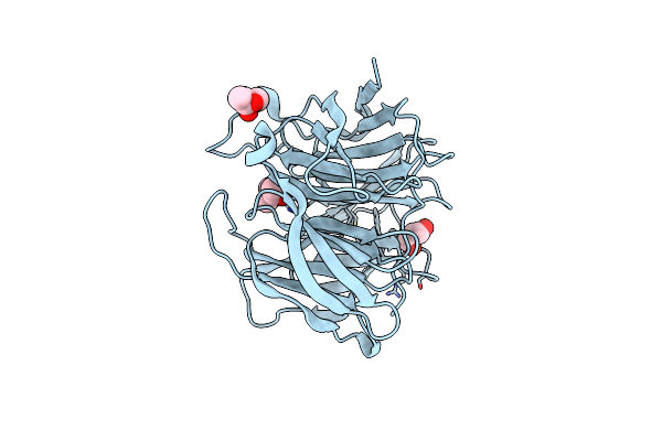
Room Temperature Rsegfp2 With A Chlorinated Chromophore 300 Fs After Photoexcitation
Organism: Aequorea victoria
Method: X-RAY DIFFRACTION Resolution:1.63 Å Release Date: 2023-07-19 Classification: FLUORESCENT PROTEIN |
 |
Room Temperature Rsegfp2 With A Chlorinated Chromophore 600 Fs After Photoexcitation
Organism: Aequorea victoria
Method: X-RAY DIFFRACTION Resolution:1.63 Å Release Date: 2023-07-19 Classification: FLUORESCENT PROTEIN |
 |
Room Temperature Rsegfp2 With A Chlorinated Chromophore 900 Fs After Photoexcitation
Organism: Aequorea victoria
Method: X-RAY DIFFRACTION Resolution:1.63 Å Release Date: 2023-07-19 Classification: FLUORESCENT PROTEIN |
 |
Room Temperature Rsegfp2 With A Chlorinated Chromophore 5 Ps After Photoexcitation
Organism: Aequorea victoria
Method: X-RAY DIFFRACTION Resolution:1.63 Å Release Date: 2023-07-19 Classification: FLUORESCENT PROTEIN |
 |
Room Temperature Rsegfp2 With A Chlorinated Chromophore 100 Ps After Photoexcitation
Organism: Aequorea victoria
Method: X-RAY DIFFRACTION Resolution:1.63 Å Release Date: 2023-07-19 Classification: FLUORESCENT PROTEIN |
 |
Room Temperature Rsegfp2 With A Chlorinated Chromophore 1 Microsecond After Photoexcitation
Organism: Aequorea victoria
Method: X-RAY DIFFRACTION Resolution:1.63 Å Release Date: 2023-07-19 Classification: FLUORESCENT PROTEIN |
 |
Organism: Aequorea victoria
Method: X-RAY DIFFRACTION Resolution:1.46 Å Release Date: 2023-07-19 Classification: FLUORESCENT PROTEIN |
 |
Rsegfp2 With A Chlorinated Chromophore In The Fluorescent On-State In A Crystal Dehydrated After Illumination
Organism: Aequorea victoria
Method: X-RAY DIFFRACTION Resolution:1.81 Å Release Date: 2023-07-19 Classification: FLUORESCENT PROTEIN Ligands: SO4 |
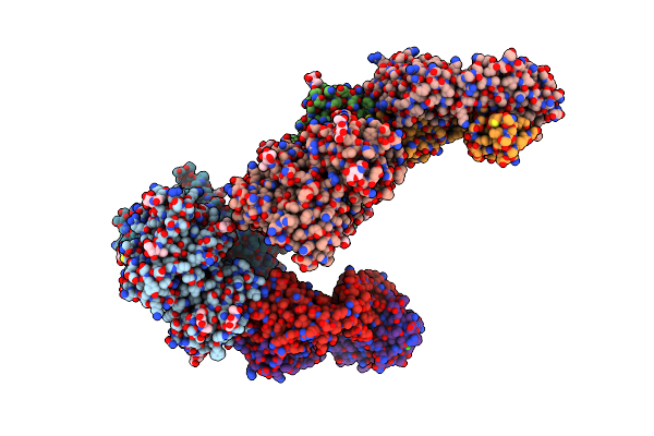 |
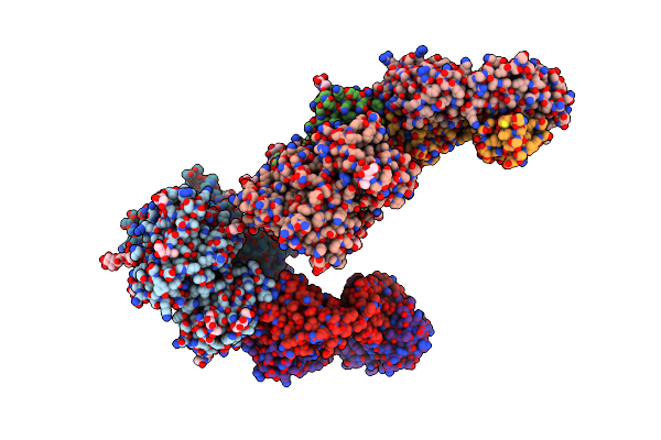
Crystal Structure Of The Medaka Fish Taste Receptor T1R2A-T1R3 Ligand Binding Domains In Complex With L-Glutamine
Organism: Oryzias latipes, Mus musculus
Method: X-RAY DIFFRACTION Resolution:2.21 Å Release Date: 2017-05-24 Classification: SIGNALING PROTEIN/IMMUNE SYSTEM Ligands: NAG, GLN, NA, CL, CA |
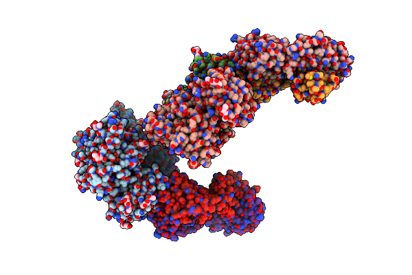 |
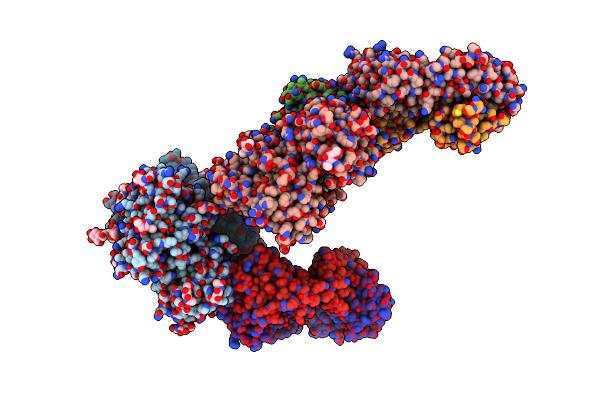
Crystal Structure Of The Medaka Fish Taste Receptor T1R2A-T1R3 Ligand Binding Domains In Complex With L-Alanine
Organism: Oryzias latipes, Mus musculus
Method: X-RAY DIFFRACTION Resolution:2.20 Å Release Date: 2017-05-24 Classification: SIGNALING PROTEIN/IMMUNE SYSTEM Ligands: NAG, ALA, NA, CL |
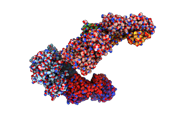 |
Crystal Structure Of The Medaka Fish Taste Receptor T1R2A-T1R3 Ligand Binding Domains In Complex With L-Arginine
Organism: Oryzias latipes, Mus musculus
Method: X-RAY DIFFRACTION Resolution:2.60 Å Release Date: 2017-05-24 Classification: SIGNALING PROTEIN/IMMUNE SYSTEM Ligands: NAG, ARG, NA, CL, CA |
 |
Crystal Structure Of The Medaka Fish Taste Receptor T1R2A-T1R3 Ligand Binding Domains In Complex With L-Glutamate
Organism: Oryzias latipes, Mus musculus
Method: X-RAY DIFFRACTION Resolution:2.61 Å Release Date: 2017-05-24 Classification: SIGNALING PROTEIN/IMMUNE SYSTEM Ligands: NAG, GLU, NA, CL, CA |
 |
Crystal Structure Of The Medaka Fish Taste Receptor T1R2A-T1R3 Ligand Binding Domains In Complex With Glycine
Organism: Oryzias latipes, Mus musculus
Method: X-RAY DIFFRACTION Resolution:2.60 Å Release Date: 2017-05-24 Classification: SIGNALING PROTEIN/IMMUNE SYSTEM Ligands: NAG, GLY, NA, CL, CA |
 |
Luciferin-Regenerating Enzyme Collected With Serial Synchrotron Rotational Crystallography With Accumulated Dose Of 1.1 Mgy (1St Measurement)
Organism: Photinus pyralis
Method: X-RAY DIFFRACTION Resolution:1.60 Å Release Date: 2017-01-04 Classification: HYDROLASE Ligands: HG, MG, MPD, GOL |
 |
Luciferin-Regenerating Enzyme Collected With Serial Synchrotron Rotational Crystallography With Accumulated Dose Of 3.4 Mgy (3Rd Measurement)
Organism: Photinus pyralis
Method: X-RAY DIFFRACTION Resolution:1.60 Å Release Date: 2017-01-04 Classification: HYDROLASE Ligands: HG, MG, MPD, GOL |
 |
Luciferin-Regenerating Enzyme Collected With Serial Synchrotron Rotational Crystallography With Accumulated Dose Of 6.9 Mgy (6Th Measurement)
Organism: Photinus pyralis
Method: X-RAY DIFFRACTION Resolution:1.60 Å Release Date: 2017-01-04 Classification: HYDROLASE Ligands: HG, MG, MPD, GOL |
 |
Luciferin-Regenerating Enzyme Collected With Serial Synchrotron Rotational Crystallography With Accumulated Dose Of 14 Mgy (12Th Measurement)
Organism: Photinus pyralis
Method: X-RAY DIFFRACTION Resolution:1.60 Å Release Date: 2017-01-04 Classification: HYDROLASE Ligands: HG, MG, MPD, GOL |