
Search Count: 25
 |
Organism: Homo sapiens
Method: X-RAY DIFFRACTION Resolution:2.60 Å Release Date: 2023-03-22 Classification: TRANSCRIPTION/INHIBITOR Ligands: 7IX, UNX, DMS |
 |
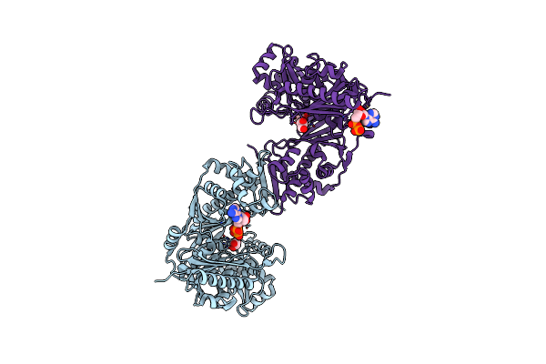
Crystal Structure Of Trypanosoma Brucei Gambiense Glycerol Kinase Complex With Amp-Pnp.
Organism: Trypanosoma brucei gambiense
Method: X-RAY DIFFRACTION Resolution:2.70 Å Release Date: 2020-01-29 Classification: TRANSFERASE Ligands: GOL, ANP |
 |
Crystal Structure Of Trypanosoma Brucei Gambiense Glycerol Kinase Complex With Adp.
Organism: Trypanosoma brucei gambiense
Method: X-RAY DIFFRACTION Resolution:2.40 Å Release Date: 2020-01-29 Classification: TRANSFERASE Ligands: ADP, GOL |
 |
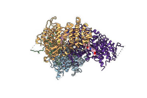
Crystal Structure Of Pyrophosphate-Dependent Phosphoenolpyruvate Carboxykinase (Ppi-Pepck)
Organism: Actinomyces israelii
Method: X-RAY DIFFRACTION Resolution:2.60 Å Release Date: 2019-11-06 Classification: LYASE Ligands: CO |
 |
Organism: Homo sapiens
Method: X-RAY DIFFRACTION Resolution:1.90 Å Release Date: 2019-09-11 Classification: MEMBRANE PROTEIN Ligands: SO4, ACT, FMN, ORO, 9AU |
 |
Organism: Eimeria tenella
Method: X-RAY DIFFRACTION Resolution:3.50 Å Release Date: 2019-08-28 Classification: MEMBRANE PROTEIN Ligands: FMN, ORO |
 |
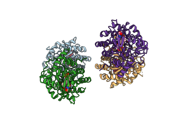
Crystal Structure Of Trypanosoma Brucei Glycosomal Isocitrate Dehydrogenase In Complex With Nadp+
Organism: Trypanosoma brucei brucei
Method: X-RAY DIFFRACTION Resolution:3.20 Å Release Date: 2019-08-28 Classification: OXIDOREDUCTASE Ligands: NAP |
 |
Crystal Structure Of Trypanosoma Brucei Glycosomal Isocitrate Dehydrogenase In Complex With Nadp+, Alpha-Ketoglutarate And Ca2+
Organism: Trypanosoma brucei brucei
Method: X-RAY DIFFRACTION Resolution:2.40 Å Release Date: 2019-08-28 Classification: OXIDOREDUCTASE Ligands: NAP, CA, AKG |
 |
Crystal Structure Of Trypanosoma Brucei Glycosomal Isocitrate Dehydrogenase In Complex With Nadph, Alpha-Ketoglutarate And Ca2+
Organism: Trypanosoma brucei brucei
Method: X-RAY DIFFRACTION Resolution:2.85 Å Release Date: 2019-08-28 Classification: OXIDOREDUCTASE Ligands: NAP, CA, GOL, AKG |
 |
Crystal Structure Of Trypanosoma Brucei Glycosomal Isocitrate Dehydrogenase In Complex With Nadh, Alpha-Ketoglutarate And Ca2+
Organism: Trypanosoma brucei brucei
Method: X-RAY DIFFRACTION Resolution:2.90 Å Release Date: 2019-08-28 Classification: OXIDOREDUCTASE Ligands: NAD, CA, AKG |
 |
Crystal Structure Of Trypanosoma Cruzi Cytosolic Isocitrate Dehydrogenase In Complex With Nadp+, Isocitrate And Ca2+
Organism: Trypanosoma cruzi (strain cl brener)
Method: X-RAY DIFFRACTION Resolution:2.40 Å Release Date: 2019-08-28 Classification: OXIDOREDUCTASE Ligands: NAP, CA, ICT |
 |
Organism: Eimeria tenella
Method: X-RAY DIFFRACTION Resolution:3.65 Å Release Date: 2019-08-28 Classification: MEMBRANE PROTEIN Ligands: FMN, ORO, 9AU |
 |
Crystal Structure Of Wild Type Vps29 Complexed With Zn+2 From Entamoeba Histolytica
Organism: Entamoeba histolytica
Method: X-RAY DIFFRACTION Resolution:2.85 Å Release Date: 2017-10-18 Classification: PROTEIN TRANSPORT Ligands: ZN |
 |
Organism: Entamoeba histolytica
Method: X-RAY DIFFRACTION Resolution:1.86 Å Release Date: 2017-10-18 Classification: PROTEIN TRANSPORT |
 |
Crystal Structure Of Vps29 Double Mutant (D62A/H86A) From Entamoeba Histolytica
Organism: Entamoeba histolytica
Method: X-RAY DIFFRACTION Resolution:2.20 Å Release Date: 2017-10-18 Classification: PROTEIN TRANSPORT |
 |
Organism: Entamoeba histolytica
Method: X-RAY DIFFRACTION Resolution:1.86 Å Release Date: 2017-10-04 Classification: TRANSPORT PROTEIN |
 |
Crystal Structure Of The Gdp-Bound Fast Hydrolyzing Mutant (V71A/K73Q) Of Ehrabx3 From Entamoeba Histolytica
Organism: Entamoeba histolytica
Method: X-RAY DIFFRACTION Resolution:3.10 Å Release Date: 2016-04-27 Classification: HYDROLASE Ligands: GDP |
 |
Crystal Structure Of The Gtp-Bound Wild Type Ehrabx3 From Entamoeba Histolytica
Organism: Entamoeba histolytica
Method: X-RAY DIFFRACTION Resolution:2.80 Å Release Date: 2016-04-27 Classification: HYDROLASE Ligands: GTP, MG |
 |
Organism: Entamoeba histolytica
Method: X-RAY DIFFRACTION Resolution:1.97 Å Release Date: 2011-02-09 Classification: LYASE Ligands: SO4, GOL |
 |
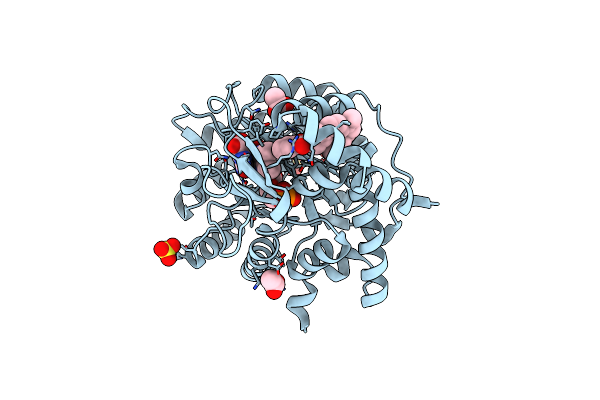
Reaction Intermediate Structure Of Entamoeba Histolytica Methionine Gamma-Lyase 1 Tetramer Containing Michaelis Complex And Methionine-Pyridoxal-5'-Phosphate
Organism: Entamoeba histolytica
Method: X-RAY DIFFRACTION Resolution:2.59 Å Release Date: 2011-02-09 Classification: LYASE Ligands: AA5, SO4, GOL, MET |