
Search Count: 75
 |
Organism: Escherichia coli
Method: ELECTRON MICROSCOPY Release Date: 2023-04-12 Classification: RIBOSOME Ligands: MG |
 |
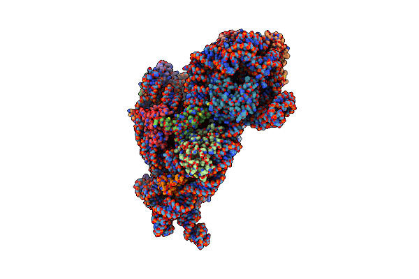
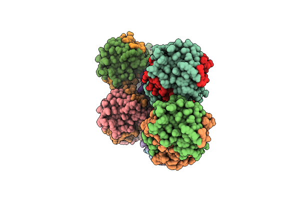
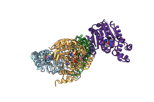
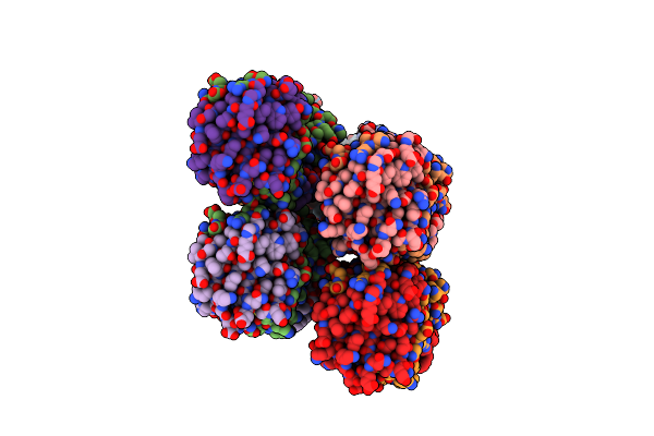
Crystal Structure Of 30S Ribosomal A1408 Methyltransferase From An Uncultured Bacterium (Unckam)
Organism: Uncultured bacterium
Method: X-RAY DIFFRACTION Resolution:1.64 Å Release Date: 2021-08-04 Classification: RNA BINDING PROTEIN Ligands: SO4 |
 |
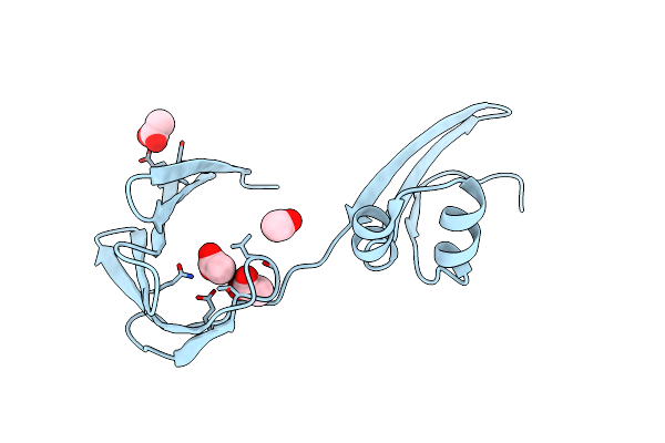
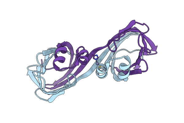
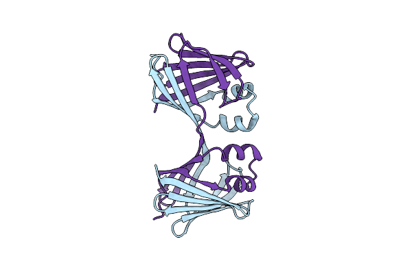
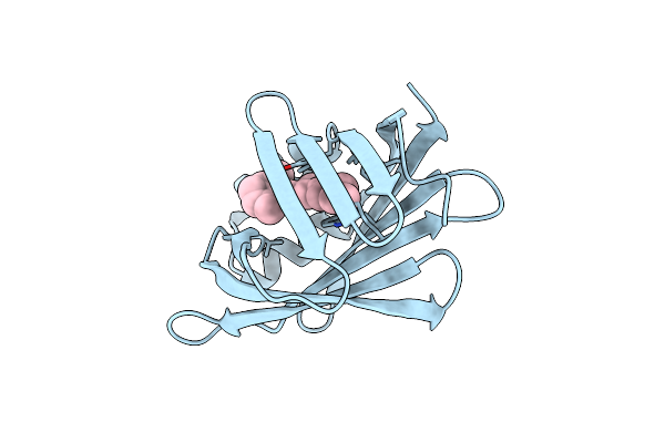
Crystal Structure Of The Apo Domain-Swapped Dimer Q108K:K40L:T51F Mutant Of Human Cellular Retinol Binding Protein Ii
Organism: Homo sapiens
Method: X-RAY DIFFRACTION Resolution:1.97 Å Release Date: 2019-10-16 Classification: CYTOSOLIC PROTEIN Ligands: ACT |
 |
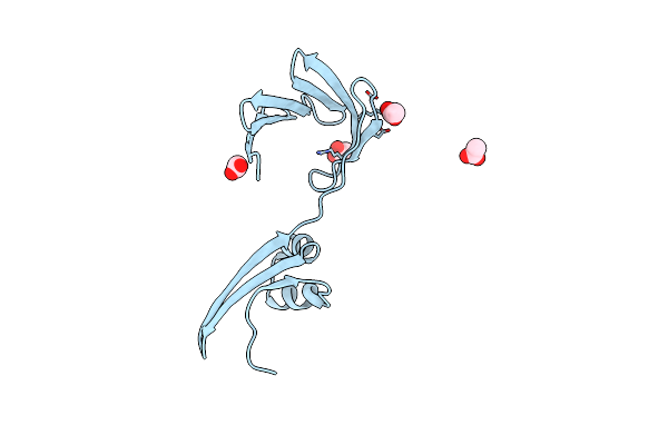
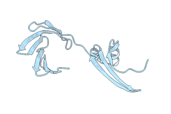
Crystal Structure Of The Apo Domain-Swapped Dimer Q108K:K40L:T51W Mutant Of Human Cellular Retinol Binding Protein Ii
Organism: Homo sapiens
Method: X-RAY DIFFRACTION Resolution:2.26 Å Release Date: 2019-10-16 Classification: LIPID BINDING PROTEIN Ligands: HOH |
 |
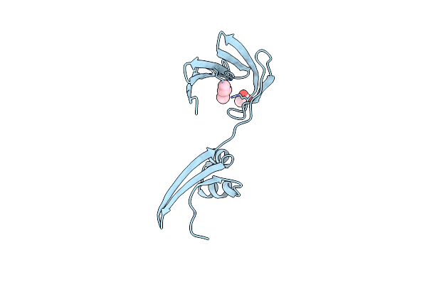
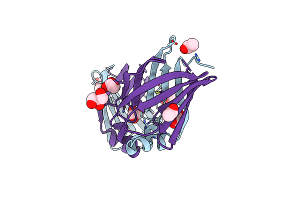
Crystal Structure Of The Apo Domain-Swapped Dimer Q108K:T51D Mutant Of Human Cellular Retinol Binding Protein Ii
Organism: Homo sapiens
Method: X-RAY DIFFRACTION Resolution:1.70 Å Release Date: 2019-10-16 Classification: LIPID BINDING PROTEIN Ligands: ACT |
 |
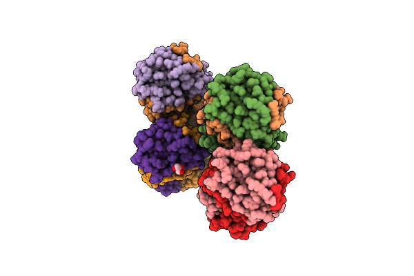
Crystal Structure Of The Apo Domain-Swapped Dimer Q108K:T51D:A28H Mutant Of Human Cellular Retinol Binding Protein Ii
Organism: Homo sapiens
Method: X-RAY DIFFRACTION Resolution:1.99 Å Release Date: 2019-10-16 Classification: LIPID BINDING PROTEIN Ligands: ACT |
 |
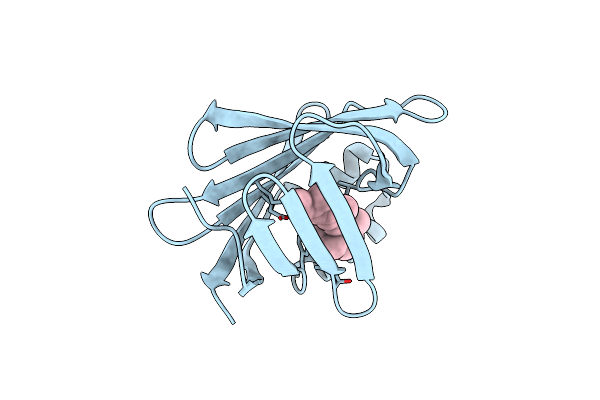
Crystal Structure Of The Holo Retinal-Bound Domain-Swapped Dimer Q108K:K40L:T51F:Y60A Mutant Of Human Cellular Retinol Binding Protein Ii
Organism: Homo sapiens
Method: X-RAY DIFFRACTION Resolution:2.08 Å Release Date: 2019-10-16 Classification: LIPID BINDING PROTEIN Ligands: ACT, RET |
 |
Crystal Structure Of The Holo Retinal-Bound Domain-Swapped Dimer Q108K:T51D:A28C Mutant Of Human Cellular Retinol Binding Protein Ii
Organism: Homo sapiens
Method: X-RAY DIFFRACTION Resolution:2.70 Å Release Date: 2019-10-16 Classification: LIPID BINDING PROTEIN Ligands: ACT, GOL, RET |
 |
Crystal Structure Of The Holo Retinal-Bound Domain-Swapped Dimer Q108K:T51D:A28H Mutant Of Human Cellular Retinol Binding Protein Ii
Organism: Homo sapiens
Method: X-RAY DIFFRACTION Resolution:2.57 Å Release Date: 2019-10-16 Classification: LIPID BINDING PROTEIN Ligands: GOL, RET |
 |
Crystal Structure Of Holo Retinal-Bound Domain-Swapped Dimer Of Wild Type Human Cellular Retinol Binding Protein Ii
Organism: Homo sapiens
Method: X-RAY DIFFRACTION Resolution:3.30 Å Release Date: 2019-10-16 Classification: LIPID BINDING PROTEIN Ligands: RET |
 |
Crystal Structure Of Retinal-Bound Holo Q108K:K40L:T51W Domain-Swapped Dimer Of Human Cellular Retinol Binding Protein 2
Organism: Homo sapiens
Method: X-RAY DIFFRACTION Resolution:2.11 Å Release Date: 2019-10-16 Classification: LIPID BINDING PROTEIN Ligands: RET, ACT, GOL |
 |
Crystal Structure Of The Holo Retinal-Bound Domain-Swapped Dimer Q108K:K40L:T51F Mutant Of Human Cellular Retinol Binding Protein Ii
Organism: Homo sapiens
Method: X-RAY DIFFRACTION Resolution:2.15 Å Release Date: 2019-10-16 Classification: LIPID BINDING PROTEIN Ligands: GOL, ACT, RET |
 |
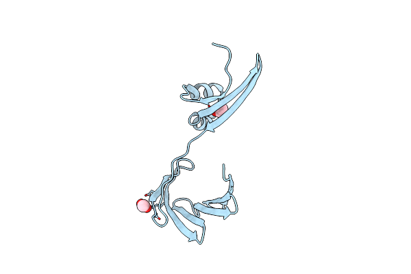
Crystal Structure Of The Zn-Bound Domain-Swapped Dimer Q108K:T51D:A28C:L36C:F57H Mutant Of Human Cellular Retinol Binding Protein Ii
Organism: Homo sapiens
Method: X-RAY DIFFRACTION Resolution:1.64 Å Release Date: 2019-10-16 Classification: LIPID BINDING PROTEIN Ligands: ZN |
 |
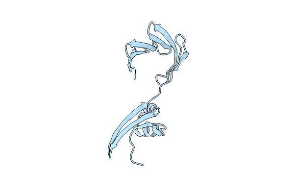
Crystal Structure Of Apo Domain-Swapped Dimer Q108K:T51D:A28C:L36C Mutant Of Human Cellular Retinol Binding Protein Ii
Organism: Homo sapiens
Method: X-RAY DIFFRACTION Resolution:1.98 Å Release Date: 2019-10-16 Classification: LIPID BINDING PROTEIN |
 |
Crystal Structure Of The Reduced Form Of Apo Domain-Swapped Dimer Q108K:T51D:A28C:L36C:F57H Mutant Of Human Cellular Retinol Binding Protein Ii
Organism: Homo sapiens
Method: X-RAY DIFFRACTION Resolution:2.40 Å Release Date: 2019-10-16 Classification: LIPID BINDING PROTEIN |
 |
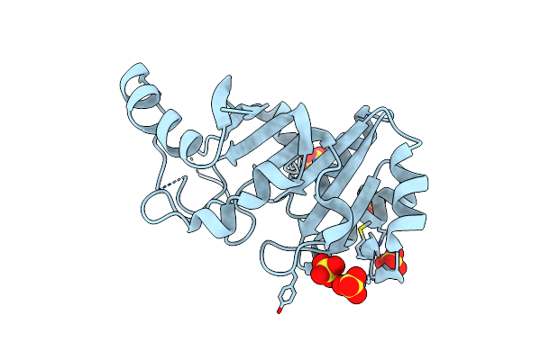
Crystal Structure Of Aminoglycoside-Resistance Methyltransferase Rmtc Bound To S-Adenosylhomocysteine (Sah)
Organism: Proteus mirabilis
Method: X-RAY DIFFRACTION Resolution:3.14 Å Release Date: 2019-10-16 Classification: TRANSFERASE Ligands: SAH |
 |
Crystal Structure Of The Apo Domain-Swapped Dimer Q108K:T51D:A28C Mutant Of Human Cellular Retinol Binding Protein Ii
Organism: Homo sapiens
Method: X-RAY DIFFRACTION Resolution:2.59 Å Release Date: 2019-08-07 Classification: LIPID BINDING PROTEIN Ligands: ACT, GOL |
 |
Crystal Structure Of Holo Retinal-Bound Domain-Swapped Dimer Q108K:T51D Mutant Of Human Cellular Retinol Binding Protein Ii
Organism: Homo sapiens
Method: X-RAY DIFFRACTION Resolution:2.06 Å Release Date: 2019-07-24 Classification: LIPID BINDING PROTEIN Ligands: RET |
 |
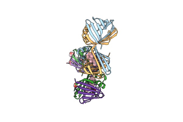
Crystal Structure Of The All-Trans Retinal-Bound R111K:Y134F:T54V:R132Q:P39Y:R59Y:L121E Mutant Of Human Cellular Retinoic Acid Binding Protein Ii In The Dark At 1.9 Angstrom Resolution
Organism: Homo sapiens
Method: X-RAY DIFFRACTION Resolution:1.90 Å Release Date: 2019-01-09 Classification: LIPID BINDING PROTEIN Ligands: RET |
 |
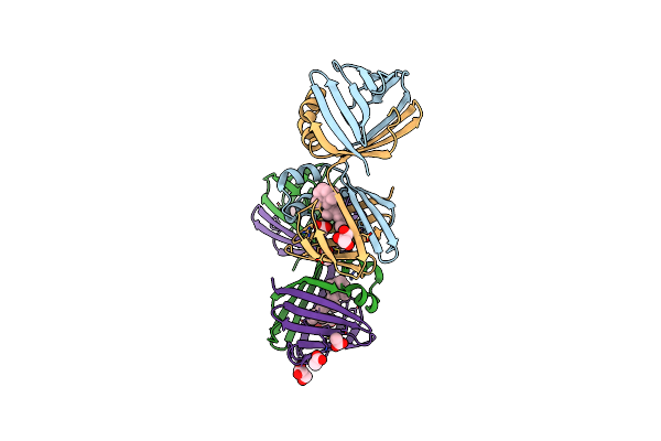
Crystal Structure Of The All-Trans Retinal-Bound R111K:Y134F:T54V:R132Q:P39Y:R59Y:L121W Mutant Of Human Cellular Retinoic Acid Binding Protein Ii In The Dark At 2.2 Angstrom Resolution
Organism: Homo sapiens
Method: X-RAY DIFFRACTION Resolution:2.20 Å Release Date: 2019-01-09 Classification: LIPID BINDING PROTEIN Ligands: RET |