
Search Count: 135
 |
 |
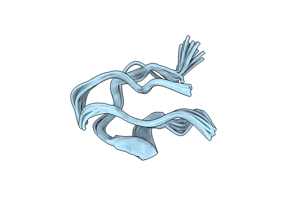
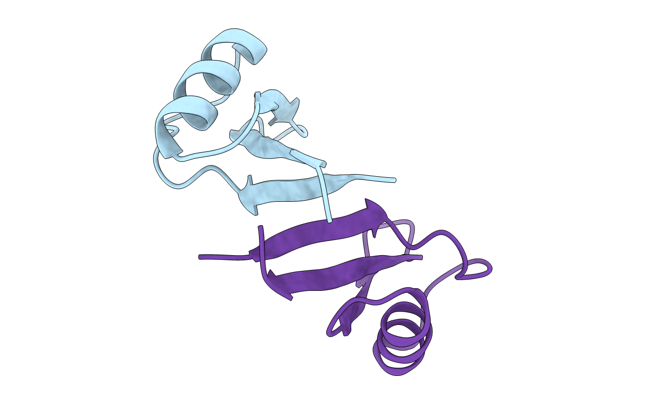
Structure Of A New Shkt Peptide From The Sea Anemone Telmatactis Stephensoni: Shkt-Ts1
Organism: Telmatactis stephensoni
Method: SOLUTION NMR Release Date: 2023-10-18 Classification: TOXIN |
 |
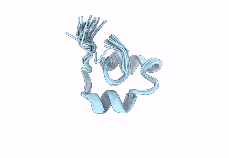
Structural And Functional Characterisation Of Tst2, A Novel Trpv1 Inhibitory Peptide From The Australian Sea Anemone Telmatactis Stephensoni
Organism: Telmatactis stephensoni
Method: SOLUTION NMR Release Date: 2023-09-27 Classification: TOXIN |
 |
Structure Of Elevenin-Vc1 From Venom Of The Australian Cone Snail Conus Victoriae
|
 |
Solution Structure Of Peptide Toxin Miitx2-Mg1A From The Venom Of The Australian Giant Red Bull Ant Myrmecia Gulosa
|
 |
 |
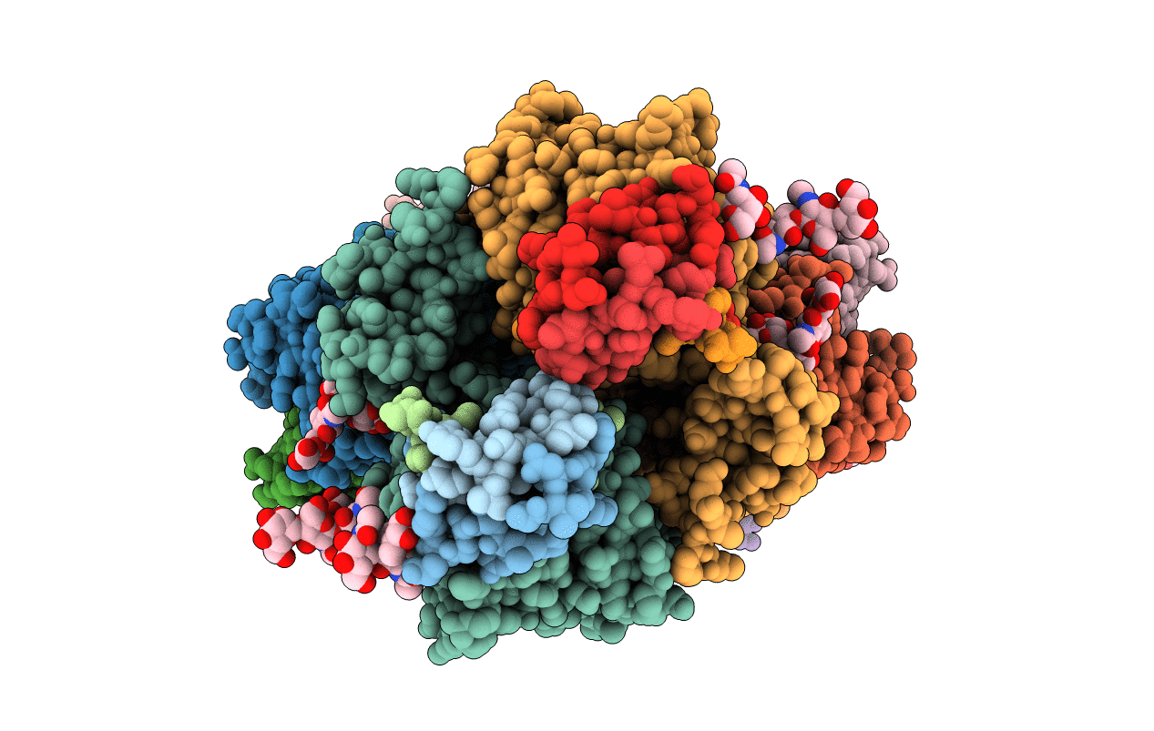
Organism: Mycobacterium tuberculosis (strain cdc 1551 / oshkosh)
Method: X-RAY DIFFRACTION Resolution:1.68 Å Release Date: 2021-06-09 Classification: ANTIBIOTIC Ligands: DMS, GDP, 4HC |
 |
Mycobacterium Tuberculosis Ftsz-Gtp-Gamma-S In Complex With 4-Hydroxycoumarin
Organism: Mycobacterium tuberculosis (strain cdc 1551 / oshkosh)
Method: X-RAY DIFFRACTION Resolution:2.40 Å Release Date: 2021-06-09 Classification: ANTIBIOTIC Ligands: DMS, GSP, 4HC |
 |
Organism: Mycobacterium tuberculosis (strain cdc 1551 / oshkosh)
Method: X-RAY DIFFRACTION Resolution:2.03 Å Release Date: 2021-04-21 Classification: CELL CYCLE Ligands: GSP |
 |
Organism: Mycobacterium tuberculosis (strain cdc 1551 / oshkosh)
Method: X-RAY DIFFRACTION Resolution:1.70 Å Release Date: 2021-04-14 Classification: ANTIBIOTIC Ligands: GDP |
 |
 |
Organism: Vitis vinifera
Method: X-RAY DIFFRACTION Resolution:1.30 Å Release Date: 2020-09-16 Classification: ANTIMICROBIAL PROTEIN |
 |
Organism: Elaeis guineensis
Method: X-RAY DIFFRACTION Resolution:2.10 Å Release Date: 2020-09-09 Classification: PROTEIN BINDING |
 |
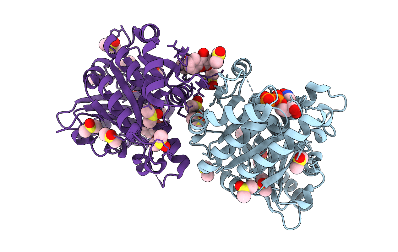
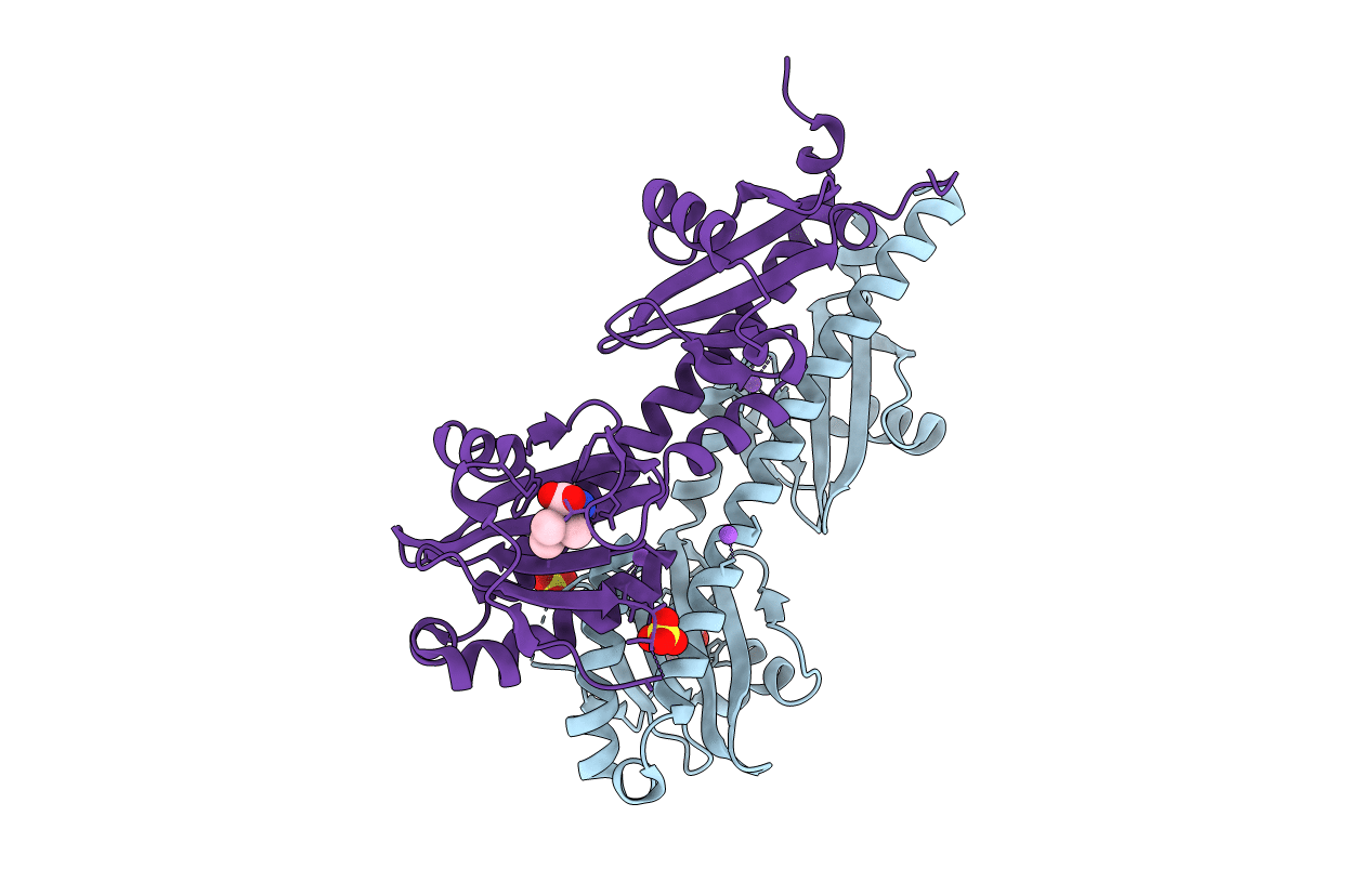
Human Insulin In Complex With The Human Insulin Microreceptor In Turn In Complex With Fv 83-7
Organism: Homo sapiens, Mus musculus
Method: X-RAY DIFFRACTION Resolution:2.90 Å Release Date: 2020-06-03 Classification: HORMONE RECEPTOR/HORMONE/IMMUNE SYSTEM Ligands: NAG |
 |
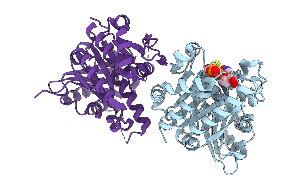
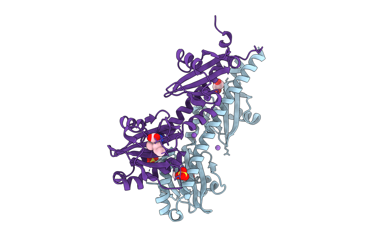
Con-Ins G1 In Complex With The Human Insulin Microreceptor In Turn In Complex With Fv 83-7
Organism: Homo sapiens, Mus musculus, Conus geographus
Method: X-RAY DIFFRACTION Resolution:3.25 Å Release Date: 2020-06-03 Classification: TOXIN Ligands: NAG, SO4 |
 |
Organism: Homo sapiens
Method: X-RAY DIFFRACTION Resolution:1.46 Å Release Date: 2020-06-03 Classification: HORMONE |
 |
Crystal Structure Of Ligand-Binding Domain Of Campylobacter Jejuni Chemoreceptor Tlp3 In Complex With 4-Methylisoleucine
Organism: Campylobacter jejuni
Method: X-RAY DIFFRACTION Resolution:1.42 Å Release Date: 2020-05-20 Classification: SIGNALING PROTEIN Ligands: SKG, SO4, NA, CL |
 |
Crystal Structure Of Ligand-Binding Domain Of Campylobacter Jejuni Chemoreceptor Tlp3 In Complex With Beta-Methylnorleucine
Organism: Campylobacter jejuni
Method: X-RAY DIFFRACTION Resolution:1.38 Å Release Date: 2020-05-20 Classification: SIGNALING PROTEIN Ligands: SKJ, SO4, CL, GOL, NA |
 |
Crystal Structure Of Ligand-Binding Domain Of Campylobacter Jejuni Chemoreceptor Tlp3 In Complex With 3-Methylisoleucine
Organism: Campylobacter jejuni
Method: X-RAY DIFFRACTION Resolution:1.38 Å Release Date: 2020-05-20 Classification: SIGNALING PROTEIN Ligands: I2M, SO4, CL, NA |
 |
Crystal Structure Of Ligand-Binding Domain Of Campylobacter Jejuni Chemoreceptor Tlp3 In Complex With L-Leucine
Organism: Campylobacter jejuni
Method: X-RAY DIFFRACTION Resolution:1.40 Å Release Date: 2020-05-20 Classification: SIGNALING PROTEIN Ligands: LEU, GOL, SO4 |