
Search Count: 19
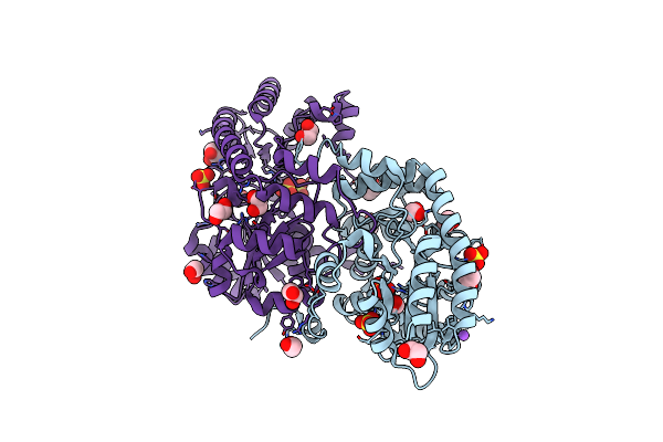 |
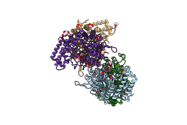
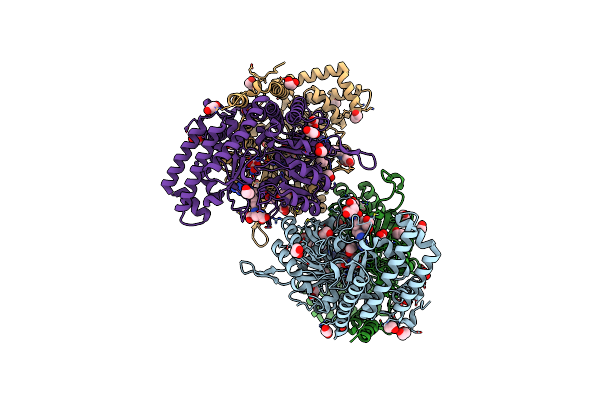
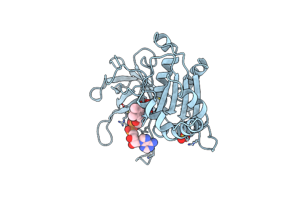
Crystal Structure Of Medicago Truncatula Histidinol-Phosphate Aminotransferase (Hisn6) In The Open State
Organism: Medicago truncatula
Method: X-RAY DIFFRACTION Resolution:1.57 Å Release Date: 2023-03-22 Classification: TRANSFERASE Ligands: SO4, EDO, NA |
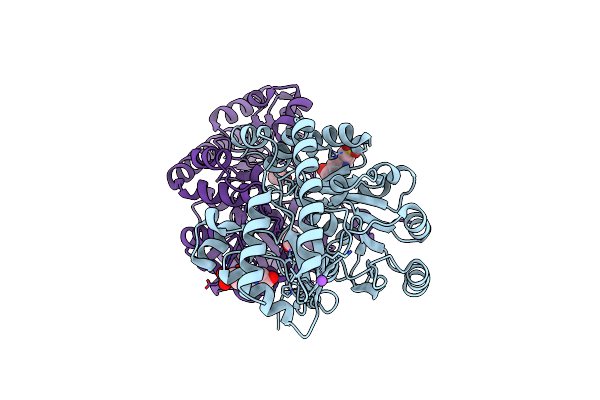 |
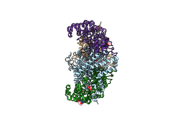
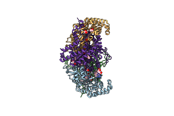
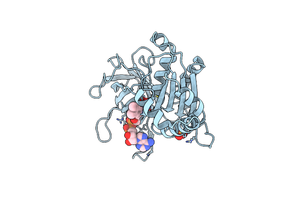
Crystal Structure Of Medicago Truncatula Histidinol-Phosphate Aminotransferase (Hisn6) In The Closed State
Organism: Medicago truncatula
Method: X-RAY DIFFRACTION Resolution:1.40 Å Release Date: 2023-03-22 Classification: TRANSFERASE Ligands: ACT, NA, EPE |
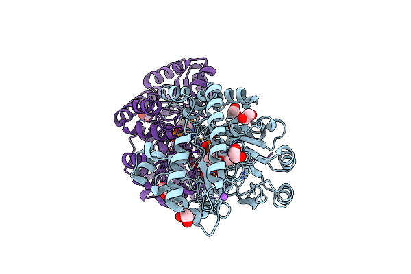 |
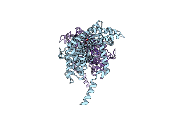
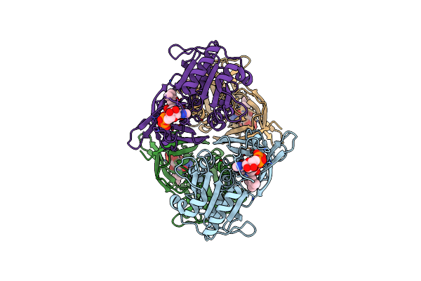
Crystal Structure Of Medicago Truncatula Histidinol-Phosphate Aminotransferase (Hisn6) In Complex With Histidinol-Phosphate
Organism: Medicago truncatula
Method: X-RAY DIFFRACTION Resolution:1.61 Å Release Date: 2023-03-22 Classification: TRANSFERASE Ligands: QNX, EDO, NA |
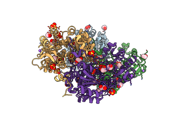 |
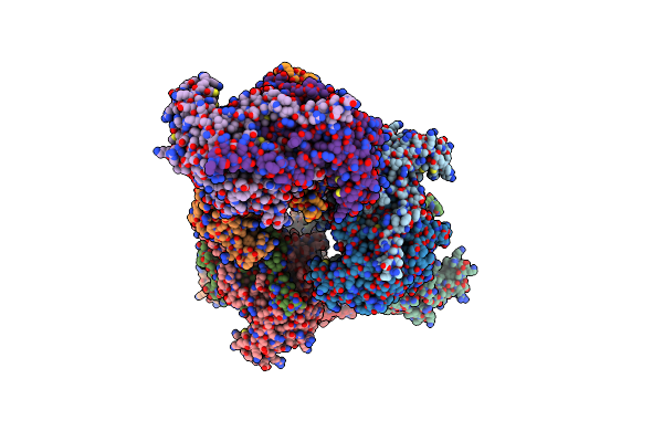
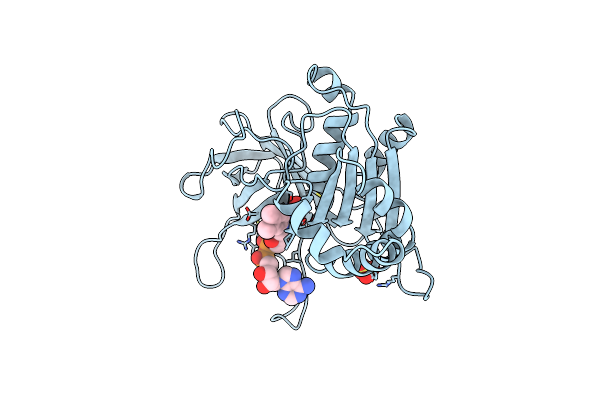
Crystal Structure Of Medicago Truncatula Histidinol-Phosphate Aminotransferase (Hisn6) In Apo Form
Organism: Medicago truncatula
Method: X-RAY DIFFRACTION Resolution:1.45 Å Release Date: 2023-03-22 Classification: TRANSFERASE Ligands: SO4, EDO, NA |
 |
Crystal Structure Of Serine Hydroxymethyltransferase, Isoform 2 From Arabidopsis Thaliana (Shm2)
Organism: Arabidopsis thaliana
Method: X-RAY DIFFRACTION Resolution:1.65 Å Release Date: 2022-08-24 Classification: TRANSFERASE Ligands: EDO, CL, EPE, PEG, SO4 |
 |
Crystal Structure Of Serine Hydroxymethyltransferase, Isoform 4 From Arabidopsis Thaliana (Shm4)
Organism: Arabidopsis thaliana
Method: X-RAY DIFFRACTION Resolution:1.74 Å Release Date: 2022-08-24 Classification: TRANSFERASE Ligands: EDO, TRS |
 |
Crystal Structure Of Serine Hydroxymethyltransferase, Isoform 6 From Arabidopsis Thaliana (Shm6)
Organism: Arabidopsis thaliana
Method: X-RAY DIFFRACTION Resolution:2.18 Å Release Date: 2022-08-24 Classification: TRANSFERASE Ligands: NO3 |
 |
Crystal Structure Of Serine Hydroxymethyltransferase, Isoform 7 From Arabidopsis Thaliana (Shm7)
Organism: Arabidopsis thaliana
Method: X-RAY DIFFRACTION Resolution:2.74 Å Release Date: 2022-08-24 Classification: TRANSFERASE |
 |
Crystal Structure Of Serine Hydroxymethyltransferase From Aphanothece Halophytica In The Covalent Complex With Malonate
Organism: Aphanothece halophytica
Method: X-RAY DIFFRACTION Resolution:1.25 Å Release Date: 2020-06-03 Classification: TRANSFERASE Ligands: PMP, EDO, MLI, NA |
 |
Crystal Structure Of Serine Hydroxymethyltransferase From Aphanothece Halophytica In The Plp-Internal Aldimine State
Organism: Aphanothece halophytica
Method: X-RAY DIFFRACTION Resolution:1.77 Å Release Date: 2020-06-03 Classification: TRANSFERASE Ligands: GOL, PEG |
 |
Crystal Structure Of Serine Hydroxymethyltransferase From Aphanothece Halophytica In The Plp-Serine External Aldimine State
Organism: Aphanothece halophytica
Method: X-RAY DIFFRACTION Resolution:1.63 Å Release Date: 2020-06-03 Classification: TRANSFERASE Ligands: GOL, PLS, 1PE |
 |
A. Thaliana Serine Hydroxymethyltransferase Isoform 2 (Atshmt2) In Complex With Methotrexate
Organism: Arabidopsis thaliana
Method: X-RAY DIFFRACTION Resolution:1.63 Å Release Date: 2020-01-08 Classification: TRANSFERASE Ligands: PLS, EDO, MTX, SER |
 |
A. Thaliana Serine Hydroxymethyltransferase Isoform 4 (Atshmt4) In Complex With Methotrexate
Organism: Arabidopsis thaliana
Method: X-RAY DIFFRACTION Resolution:2.12 Å Release Date: 2020-01-08 Classification: TRANSFERASE Ligands: PLS, EDO, MTX, TRS |
 |
A. Thaliana Serine Hydroxymethyltransferase Isoform 2 (Atshmt2) In Complex With Pemetrexed
Organism: Arabidopsis thaliana
Method: X-RAY DIFFRACTION Resolution:1.54 Å Release Date: 2020-01-08 Classification: TRANSFERASE Ligands: PLS, EDO, LYA, SER, PLP |
 |
X-Ray Structure Of The Ferredoxin-Nadp(H) Reductase From Rhodobacter Capsulatus Complexed With Three Molecules Of The Detergent N-Heptyl- Beta-D-Thioglucoside At 1.7 Angstroms
Organism: Rhodobacter capsulatus
Method: X-RAY DIFFRACTION Resolution:1.68 Å Release Date: 2005-09-07 Classification: OXIDOREDUCTASE Ligands: FAD, HTG, SO4, CO2 |
 |
X-Ray Structure Of The Ferredoxin-Nadp(H) Reductase From Rhodobacter Capsulatus At 2.1 Angstroms
Organism: Rhodobacter capsulatus
Method: X-RAY DIFFRACTION Resolution:2.10 Å Release Date: 2005-09-07 Classification: OXIDOREDUCTASE Ligands: FAD |
 |
Organism: Nostoc sp.
Method: X-RAY DIFFRACTION Resolution:2.30 Å Release Date: 2002-02-27 Classification: OXIDOREDUCTASE Ligands: SO4, FAD |
 |
Ferredoxin:Nadp+ Reductase Mutant With Leu 76 Mutated By Asp And Leu 78 Mutated By Asp
Organism: Nostoc sp.
Method: X-RAY DIFFRACTION Resolution:1.93 Å Release Date: 2002-02-27 Classification: OXIDOREDUCTASE Ligands: SO4, FAD |
 |
Organism: Nostoc sp.
Method: X-RAY DIFFRACTION Resolution:2.30 Å Release Date: 2001-11-28 Classification: OXIDOREDUCTASE Ligands: FAD, SO4 |