
Search Count: 281
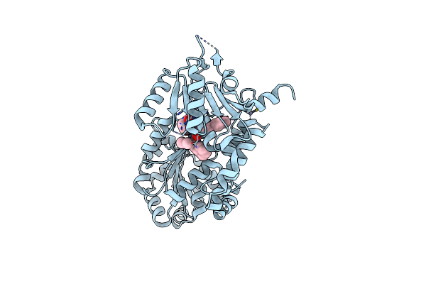 |
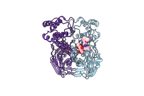
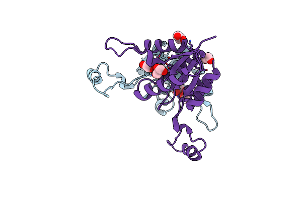
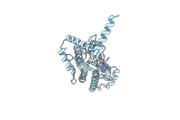
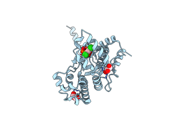
Organism: Homo sapiens
Method: X-RAY DIFFRACTION Release Date: 2025-06-18 Classification: HYDROLASE Ligands: A1CA4, ZN |
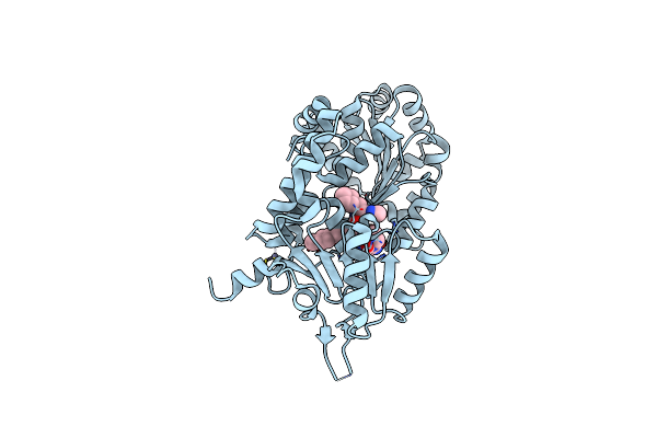 |
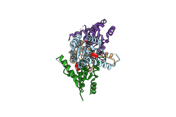
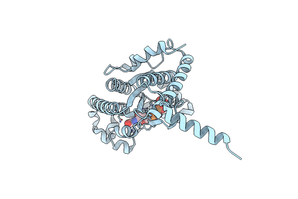
Organism: Homo sapiens
Method: X-RAY DIFFRACTION Release Date: 2025-06-18 Classification: HYDROLASE Ligands: A1CA5, ZN |
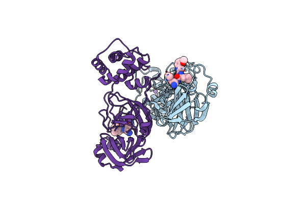 |
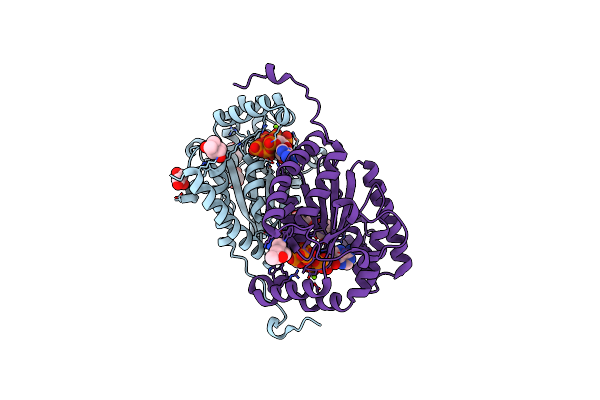
Organism: Severe acute respiratory syndrome coronavirus 2
Method: X-RAY DIFFRACTION Release Date: 2025-04-30 Classification: VIRAL PROTEIN, HYDROLASE/INHIBITOR Ligands: A1BCZ, NA |
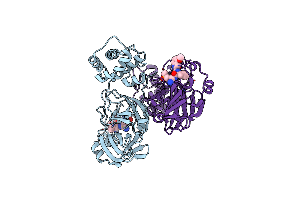 |
Organism: Severe acute respiratory syndrome coronavirus 2
Method: X-RAY DIFFRACTION Release Date: 2025-04-30 Classification: VIRAL PROTEIN, HYDROLASE/INHIBITOR Ligands: A1BCY |
 |
Organism: Severe acute respiratory syndrome coronavirus 2
Method: X-RAY DIFFRACTION Release Date: 2025-04-30 Classification: VIRAL PROTEIN, HYDROLASE/INHIBITOR Ligands: A1BCX |
 |
Organism: Severe acute respiratory syndrome coronavirus 2
Method: X-RAY DIFFRACTION Release Date: 2025-04-30 Classification: VIRAL PROTEIN, HYDROLASE/INHIBITOR Ligands: A1BCW |
 |
Crystal Structure Of A 2`-Deoxyribosyltransferase From The Psychrophilic Bacterium Desulfotalea Psychrophila.
Organism: Desulfotalea psychrophila (strain lsv54 / dsm 12343)
Method: X-RAY DIFFRACTION Resolution:2.40 Å Release Date: 2021-10-20 Classification: TRANSFERASE Ligands: GOL |
 |
The Crystal Structure From Microfluidic Crystals Of Glycosyl Hydrolase Family 2 (Gh2) Member From Bacteroides Cellulosilyticus
Organism: Bacteroides cellulosilyticus dsm 14838
Method: X-RAY DIFFRACTION Resolution:2.40 Å Release Date: 2021-08-25 Classification: HYDROLASE |
 |
Organism: Homo sapiens
Method: X-RAY DIFFRACTION Resolution:2.70 Å Release Date: 2021-07-21 Classification: SUGAR BINDING PROTEIN Ligands: M6P, ACT |
 |
Organism: Homo sapiens
Method: ELECTRON MICROSCOPY Release Date: 2021-07-14 Classification: TRANSLATION Ligands: F6P |
 |
Organism: Enterococcus faecalis
Method: X-RAY DIFFRACTION Resolution:2.15 Å Release Date: 2021-04-07 Classification: TRANSFERASE Ligands: GOL, EDO |
 |
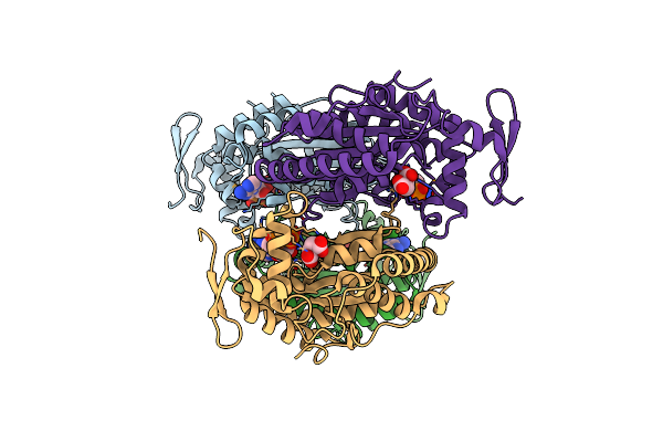
Organism: Rattus norvegicus, Gallus gallus, Sus scrofa
Method: X-RAY DIFFRACTION Resolution:3.34 Å Release Date: 2021-03-31 Classification: LIGASE Ligands: GTP, MG, CA, GDP, MES, O9B, O9H, OH5, PGE, VAL, ACP |
 |
Organism: Rattus norvegicus, Gallus gallus, Sus scrofa
Method: X-RAY DIFFRACTION Resolution:2.85 Å Release Date: 2021-03-31 Classification: LIGASE Ligands: GTP, MG, CA, GDP, MES, O9B, O9K, O9N, PGE, VAL, P6S, PEG, ACP |
 |
Crystal Structure Of Polyphosphate Kinase 2 Class I (Smc02148) In Complex With Adp
Organism: Rhizobium meliloti (strain 1021)
Method: X-RAY DIFFRACTION Resolution:1.87 Å Release Date: 2019-07-10 Classification: TRANSFERASE Ligands: ADP, MLT, AMP |
 |
Crystal Structure Of Ppk2 Class Iii In Complex With Adp From Cytophaga Hutchinsonii Atcc 33406
Organism: Cytophaga hutchinsonii (strain atcc 33406 / ncimb 9469)
Method: X-RAY DIFFRACTION Resolution:1.89 Å Release Date: 2019-01-16 Classification: TRANSFERASE Ligands: ADP, GOL |
 |
Crystal Structure Of Ppk2 Class Iii In The Complex With Amp From Cytophaga Hutchinsonii Atcc 33406
Organism: Cytophaga hutchinsonii (strain atcc 33406 / ncimb 9469)
Method: X-RAY DIFFRACTION Resolution:2.45 Å Release Date: 2019-01-16 Classification: TRANSFERASE Ligands: AMP, CL |
 |
Crystal Structure Of Ppk2 Class Iii In Complex With Guanosine 5-Tetraphosphate
Organism: Cytophaga hutchinsonii (strain atcc 33406 / ncimb 9469)
Method: X-RAY DIFFRACTION Resolution:2.65 Å Release Date: 2019-01-16 Classification: TRANSFERASE Ligands: BKP |
 |
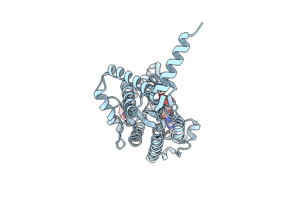
Organism: Deinococcus radiodurans (strain atcc 13939 / dsm 20539 / jcm 16871 / lmg 4051 / nbrc 15346 / ncimb 9279 / r1 / vkm b-1422)
Method: X-RAY DIFFRACTION Resolution:1.81 Å Release Date: 2019-01-16 Classification: TRANSFERASE Ligands: ATP, MG, GOL, MPD, CL |
 |
Organism: Cytophaga hutchinsonii (strain atcc 33406 / ncimb 9469)
Method: X-RAY DIFFRACTION Resolution:2.20 Å Release Date: 2019-01-16 Classification: transferase/transferase inhibitor Ligands: BOY, GOL, SRT |
 |
Crystal Structure Of Ppk2 (Class Iii) In Complex With Bisphosphonate Inhibitor (2-((3,5-Dichlorophenyl)Amino)Ethane-1,1-Diyl)Diphosphonic Acid
Organism: Cytophaga hutchinsonii (strain atcc 33406 / ncimb 9469)
Method: X-RAY DIFFRACTION Resolution:2.10 Å Release Date: 2019-01-16 Classification: transferase/transferase inhibitor Ligands: BWJ, GOL |