Planned Maintenance: Some services may turn out to be unavailable from 15th January, 2026 to 16th January, 2026. We apologize for the inconvenience!

Planned Maintenance: Some services may turn out to be unavailable from 15th January, 2026 to 16th January, 2026. We apologize for the inconvenience!
 |
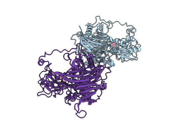
Organism: Faecalibacterium duncaniae
Method: X-RAY DIFFRACTION Release Date: 2025-12-31 Classification: TRANSFERASE Ligands: TRS, GOL, EDO, PEG |
 |
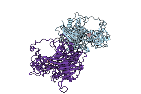
Organism: Faecalibacterium duncaniae
Method: X-RAY DIFFRACTION Release Date: 2025-12-31 Classification: TRANSFERASE Ligands: TRS, EDO |
 |
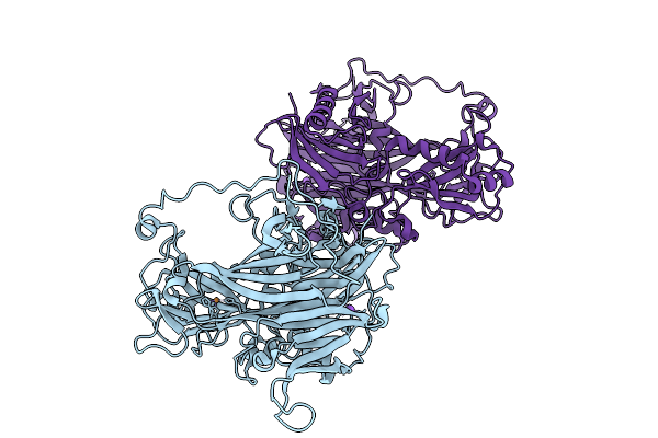
Reductive-Half Reaction Intermediate Of Copper Amine Oxidase From Arthrobacter Globiformis Captured By Mix-And-Inject Serial Crystallography At 100-Ms Time Delay
Organism: Arthrobacter globiformis
Method: X-RAY DIFFRACTION Release Date: 2025-12-31 Classification: OXIDOREDUCTASE Ligands: PEA, NA, CU |
 |
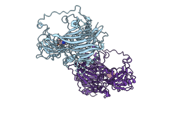
Reductive-Half Reaction Intermediate Of Copper Amine Oxidase From Arthrobacter Globiformis Captured By Mix-And-Inject Serial Crystallography At 200-Ms Time Delay
Organism: Arthrobacter globiformis
Method: X-RAY DIFFRACTION Release Date: 2025-12-31 Classification: OXIDOREDUCTASE Ligands: PEA, NA, CU |
 |
Reductive-Half Reaction Intermediate Of Copper Amine Oxidase From Arthrobacter Globiformis Captured By Mix-And-Inject Serial Crystallography At 300-Ms Time Delay
Organism: Arthrobacter globiformis
Method: X-RAY DIFFRACTION Release Date: 2025-12-31 Classification: OXIDOREDUCTASE |
 |
Reductive-Half Reaction Intermediate Of Copper Amine Oxidase From Arthrobacter Globiformis Captured By Mix-And-Inject Serial Crystallography At 400-Ms Time Delay
Organism: Arthrobacter globiformis
Method: X-RAY DIFFRACTION Release Date: 2025-12-31 Classification: OXIDOREDUCTASE Ligands: PEA, NA, CU |
 |
Reductive-Half Reaction Intermediate Of Copper Amine Oxidase From Arthrobacter Globiformis Captured With Short-A-Axis Diffraction Data By Mix-And-Inject Serial Crystallography At 22-Ms Time Delay
Organism: Arthrobacter globiformis
Method: X-RAY DIFFRACTION Release Date: 2025-12-31 Classification: OXIDOREDUCTASE Ligands: CU, NA |
 |
Reductive-Half Reaction Intermediate Of Copper Amine Oxidase From Arthrobacter Globiformis Captured With Long-A-Axis Diffraction Data By Mix-And-Inject Serial Crystallography At 22-Ms Time Delay
Organism: Arthrobacter globiformis
Method: X-RAY DIFFRACTION Release Date: 2025-12-31 Classification: OXIDOREDUCTASE Ligands: CU, NA |
 |
Reductive-Half Reaction Intermediate Of Copper Amine Oxidase From Arthrobacter Globiformis Captured With Short-A-Axis Diffraction Data By Mix-And-Inject Serial Crystallography At 25-Ms Time Delay
Organism: Arthrobacter globiformis
Method: X-RAY DIFFRACTION Release Date: 2025-12-31 Classification: OXIDOREDUCTASE Ligands: CU |
 |
Reductive-Half Reaction Intermediate Of Copper Amine Oxidase From Arthrobacter Globiformis Captured With Long-A-Axis Diffraction Data By Mix-And-Inject Serial Crystallography At 25-Ms Time Delay
Organism: Arthrobacter globiformis
Method: X-RAY DIFFRACTION Release Date: 2025-12-31 Classification: OXIDOREDUCTASE Ligands: CU, NA |
 |
Reductive-Half Reaction Intermediate Of Copper Amine Oxidase From Arthrobacter Globiformis Captured With Short-A-Axis Diffraction Data By Mix-And-Inject Serial Crystallography At 50-Ms Time Delay
Organism: Arthrobacter globiformis
Method: X-RAY DIFFRACTION Release Date: 2025-12-31 Classification: OXIDOREDUCTASE Ligands: CU |
 |
Reductive-Half Reaction Intermediate Of Copper Amine Oxidase From Arthrobacter Globiformis Captured With Long-A-Axis Diffraction Data By Mix-And-Inject Serial Crystallography At 50-Ms Time Delay
Organism: Arthrobacter globiformis
Method: X-RAY DIFFRACTION Release Date: 2025-12-31 Classification: OXIDOREDUCTASE Ligands: CU, NA |
 |
Oxidative Form Of Copper Amine Oxidase From Arthrobacter Globiformis Determined At Ph 9.0 By Single-Flow Serial Crystallography
Organism: Arthrobacter globiformis
Method: X-RAY DIFFRACTION Release Date: 2025-12-31 Classification: OXIDOREDUCTASE Ligands: CU, NA |
 |
Reductive-Half Reaction Intermediate Of Copper Amine Oxidase From Arthrobacter Globiformis Captured By Mix-And-Inject Serial Crystallography At 500-Ms Time Delay
Organism: Arthrobacter globiformis
Method: X-RAY DIFFRACTION Release Date: 2025-12-31 Classification: OXIDOREDUCTASE Ligands: CU, NA, PEA |
 |
Reductive-Half Reaction Intermediate Of Copper Amine Oxidase From Arthrobacter Globiformis Captured By Mix-And-Inject Serial Crystallography At 1000-Ms Time Delay
Organism: Arthrobacter globiformis
Method: X-RAY DIFFRACTION Release Date: 2025-12-31 Classification: OXIDOREDUCTASE Ligands: CU, NA, PEA |
 |
Organism: Homo sapiens
Method: ELECTRON MICROSCOPY Release Date: 2025-12-17 Classification: MEMBRANE PROTEIN |
 |
Organism: Bombyx mori
Method: X-RAY DIFFRACTION Release Date: 2025-12-10 Classification: PROTEIN BINDING Ligands: GDP, MG |
 |
Crystal Structure Of Keap1 In Complex With A Small Molecule Inhibitor, Kmn003
Organism: Mus musculus
Method: X-RAY DIFFRACTION Release Date: 2025-11-12 Classification: PEPTIDE BINDING PROTEIN/INHIBITOR Ligands: A1L98, FMT |
 |
Cryo-Em Structure Of Tmprss2 In Complex With Fab Fragments Of 752 Mab And 2228 Mab
Organism: Homo sapiens, Mus musculus
Method: ELECTRON MICROSCOPY Release Date: 2025-10-15 Classification: MEMBRANE PROTEIN |
 |
Organism: Homo sapiens
Method: X-RAY DIFFRACTION Release Date: 2025-10-15 Classification: IMMUNE SYSTEM Ligands: NAG |