
Search Count: 41
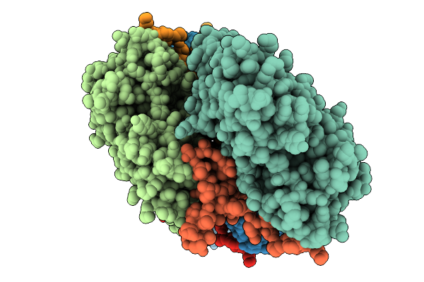 |
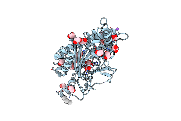
Atomic Structure Of Vibrio Effector Fragment Vopv Bound To Beta-Cytoplasmic/Gamma1-Cytoplasmic F-Actin
Organism: Homo sapiens, Vibrio parahaemolyticus
Method: ELECTRON MICROSCOPY Release Date: 2025-11-19 Classification: STRUCTURAL PROTEIN Ligands: MG, ANP |
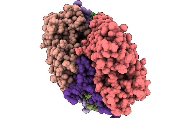 |
Cryo-Em Structure Of Vibrio Effector Vopv Fragment Bound To Skeletal Alpha F-Actin
Organism: Oryctolagus cuniculus, Vibrio cholerae
Method: ELECTRON MICROSCOPY Release Date: 2025-11-19 Classification: STRUCTURAL PROTEIN Ligands: MG, ANP |
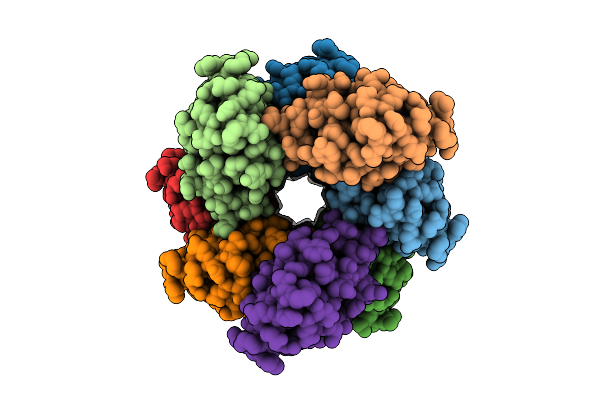 |
Organism: Homo sapiens
Method: ELECTRON MICROSCOPY Release Date: 2025-11-12 Classification: IMMUNE SYSTEM |
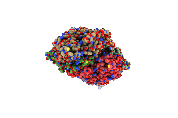 |
Organism: Salmonella enterica subsp. enterica serovar typhimurium str. lt2, Oryctolagus cuniculus
Method: ELECTRON MICROSCOPY Release Date: 2023-12-27 Classification: CELL INVASION Ligands: ADP, MG, PO4 |
 |
Organism: Homo sapiens
Method: X-RAY DIFFRACTION Resolution:2.70 Å Release Date: 2022-01-12 Classification: CELL CYCLE Ligands: ZN |
 |
Organism: Homo sapiens
Method: X-RAY DIFFRACTION Resolution:2.50 Å Release Date: 2022-01-12 Classification: CELL CYCLE Ligands: ZN |
 |
Organism: Homo sapiens
Method: X-RAY DIFFRACTION Resolution:2.60 Å Release Date: 2022-01-12 Classification: CELL CYCLE Ligands: ZN |
 |
Organism: Homo sapiens
Method: X-RAY DIFFRACTION Resolution:2.69 Å Release Date: 2022-01-12 Classification: CELL CYCLE Ligands: ZN |
 |
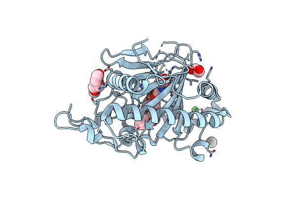
Crystal Structure Of 3-Ketosteroid Delta1-Dehydrogenase From Sterolibacterium Denitrificans In Complex With 1,4-Androstadiene-3,17-Dione
Organism: Sterolibacterium denitrificans
Method: X-RAY DIFFRACTION Resolution:1.84 Å Release Date: 2021-07-21 Classification: OXIDOREDUCTASE Ligands: FAD, ANB, PEG, GOL, NA |
 |
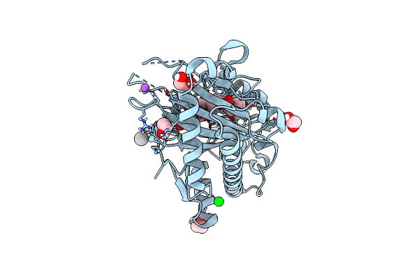
The Crystal Structure Of Kanamycin B Dioxygenase (Kanj) From Streptomyces Kanamyceticus Complex With Nickel, Sulfate And Chloride
Organism: Streptomyces kanamyceticus
Method: X-RAY DIFFRACTION Resolution:2.50 Å Release Date: 2020-07-08 Classification: ANTIBIOTIC Ligands: NI, SO4, CL |
 |
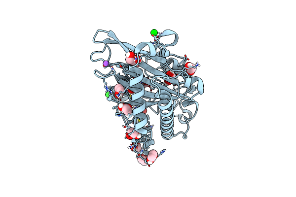
The Crystal Structure Of Kanamycin B Dioxygenase (Kanj) From Streptomyces Kanamyceticus In Complex With Nickel, Ribostamycin B And 2-Oxoglutarate
Organism: Streptomyces kanamyceticus
Method: X-RAY DIFFRACTION Resolution:2.40 Å Release Date: 2020-07-08 Classification: METAL BINDING PROTEIN Ligands: NI, AKG, RIO, CL |
 |
The Crystal Structure Of Kanamycin B Dioxygenase (Kanj) From Streptomyces Kanamyceticus In Complex With Nickel, Sulfate, Soaked With Iodide
Organism: Streptomyces kanamyceticus
Method: X-RAY DIFFRACTION Resolution:2.10 Å Release Date: 2020-07-08 Classification: METAL BINDING PROTEIN Ligands: NI, SO4, IOD |
 |
The Crystal Structure Of Kanamycin B Dioxygenase (Kanj) From Streptomyces Kanamyceticus In Complex With Nickel And 2-Oxoglutarate
Organism: Streptomyces kanamyceticus
Method: X-RAY DIFFRACTION Resolution:2.15 Å Release Date: 2020-07-08 Classification: METAL BINDING PROTEIN Ligands: AKG, NI, PEG, CL, NA |
 |
The Crystal Structure Of Kanamycin B Dioxygenase (Kanj) From Streptomyces Kanamyceticus In Complex With Nickel, Neamine And Sulfate
Organism: Streptomyces kanamyceticus
Method: X-RAY DIFFRACTION Resolution:3.00 Å Release Date: 2020-07-08 Classification: METAL BINDING PROTEIN Ligands: NI, SO4, XXX |
 |
The Crystal Structure Of Kanamycin B Dioxygenase (Kanj) From Streptomyces Kanamyceticus In Complex With Nickel And Kanamycin B Sulfate
Organism: Streptomyces kanamyceticus
Method: X-RAY DIFFRACTION Resolution:2.36 Å Release Date: 2020-07-08 Classification: METAL BINDING PROTEIN Ligands: 9CS, NI, SO4, PEG |
 |
Organism: Datura metel
Method: X-RAY DIFFRACTION Resolution:1.91 Å Release Date: 2020-03-18 Classification: OXIDOREDUCTASE Ligands: PEG, HYO, NI, OGA, EDO, SR, UNX |
 |
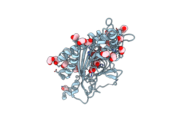
N-Terminally Truncated Hyoscyamine 6-Hydroxylase (Th6H) In Complex With N-Oxalylglycine And Hyoscyamine
Organism: Datura metel
Method: X-RAY DIFFRACTION Resolution:1.12 Å Release Date: 2020-03-18 Classification: OXIDOREDUCTASE Ligands: OGA, EDO, HYO, SR, NI, NA, UNX |
 |
N-Terminally Truncated Hyoscyamine 6-Hydroxylase (Th6H) In Complex With 2-Oxoglutarate
Organism: Datura metel
Method: X-RAY DIFFRACTION Resolution:1.31 Å Release Date: 2020-03-18 Classification: OXIDOREDUCTASE Ligands: AKG, EDO, FMT, NI, SR, NA |
 |
Thebaine 6-O-Demethylase (T6Odm) From Papaver Somniferum In Complex With Succinate
Organism: Papaver somniferum
Method: X-RAY DIFFRACTION Resolution:1.97 Å Release Date: 2018-02-14 Classification: OXIDOREDUCTASE Ligands: NI, SIN, EDO, PEG |
 |
Thebaine 6-O-Demethylase (T6Odm) From Papaver Somniferum In Complex With 2-Oxoglutarate
Organism: Papaver somniferum
Method: X-RAY DIFFRACTION Resolution:1.85 Å Release Date: 2018-02-14 Classification: OXIDOREDUCTASE Ligands: NI, AKG, EDO, UNX, PEG, NA |