
Search Count: 45
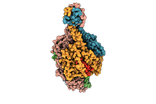 |
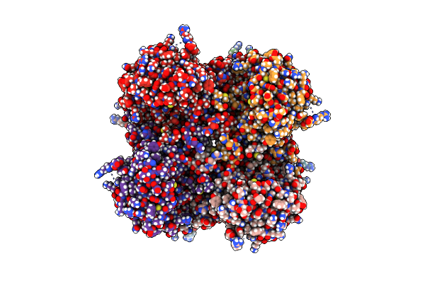
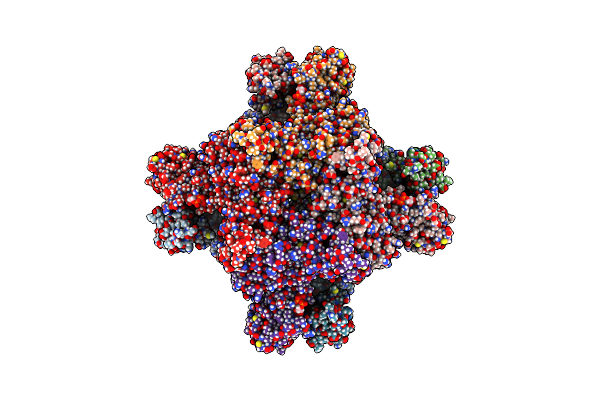
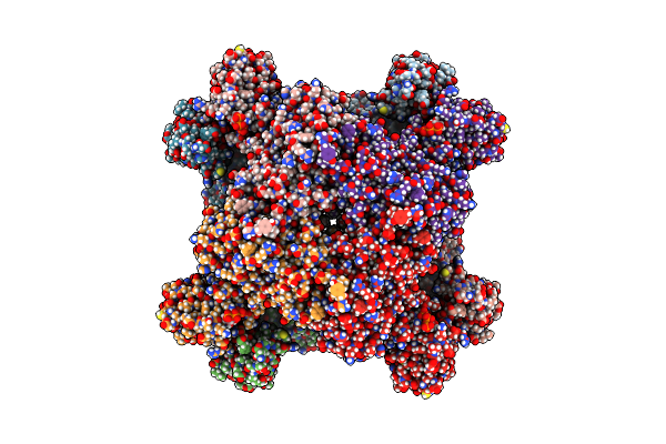
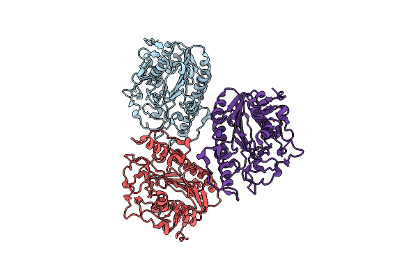
Respiratory Syncytial Virus Pre-F Trimer Bound By Neutralizing Antibody Pr306007
Organism: Respiratory syncytial virus a2, Homo sapiens
Method: ELECTRON MICROSCOPY Release Date: 2025-11-12 Classification: STRUCTURAL PROTEIN/IMMUNE SYSTEM |
 |
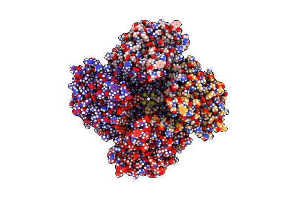
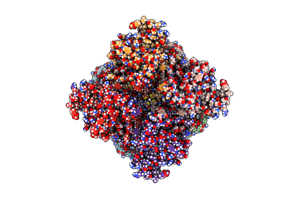
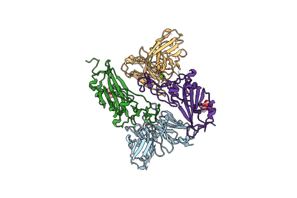
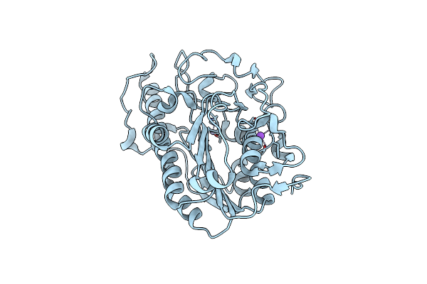
Organism: Homo sapiens
Method: X-RAY DIFFRACTION Resolution:2.73 Å Release Date: 2023-04-19 Classification: DNA BINDING PROTEIN |
 |
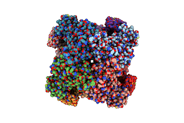
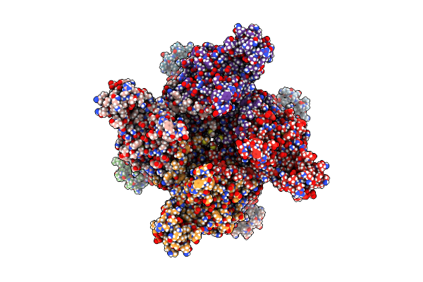
Organism: Homo sapiens
Method: ELECTRON MICROSCOPY Release Date: 2022-01-12 Classification: OXIDOREDUCTASE Ligands: NAD, IMP |
 |
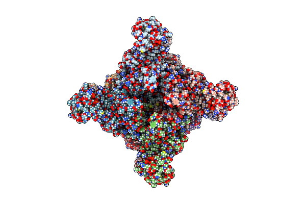
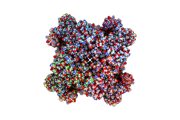
Organism: Homo sapiens
Method: ELECTRON MICROSCOPY Release Date: 2022-01-12 Classification: OXIDOREDUCTASE Ligands: ATP, IMP, NAD |
 |
Organism: Homo sapiens
Method: ELECTRON MICROSCOPY Release Date: 2022-01-12 Classification: OXIDOREDUCTASE Ligands: GTP, ATP, IMP |
 |
Organism: Homo sapiens
Method: ELECTRON MICROSCOPY Release Date: 2022-01-12 Classification: OXIDOREDUCTASE |
 |
Organism: Homo sapiens
Method: ELECTRON MICROSCOPY Release Date: 2022-01-12 Classification: OXIDOREDUCTASE Ligands: GTP, ATP, IMP |
 |
Organism: Homo sapiens
Method: ELECTRON MICROSCOPY Release Date: 2022-01-12 Classification: OXIDOREDUCTASE Ligands: ATP |
 |
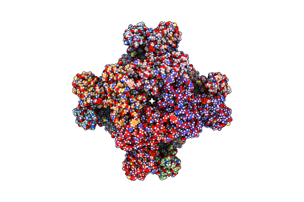
Human Retinal Variant Impdh1(595) Treated With Gtp, Atp, Imp, Nad+, Interface-Centered
Organism: Homo sapiens
Method: ELECTRON MICROSCOPY Release Date: 2022-01-12 Classification: OXIDOREDUCTASE Ligands: GTP, IMP, NAD |
 |
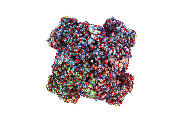
Human Retinal Variant Impdh1(595) Treated With Gtp, Atp, Imp, Nad+, Octamer-Centered
Organism: Homo sapiens
Method: ELECTRON MICROSCOPY Release Date: 2022-01-12 Classification: OXIDOREDUCTASE Ligands: GTP, ATP, IMP, NAD |
 |
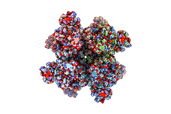
Human Retinal Variant Impdh1(546) Treated With Gtp, Atp, Imp, Nad+; Interface-Centered
Organism: Homo sapiens
Method: ELECTRON MICROSCOPY Release Date: 2022-01-12 Classification: OXIDOREDUCTASE Ligands: NAD, IMP |
 |
Human Retinal Variant Impdh1(546) Treated With Atp, Imp, Nad+, Interface-Centered
Organism: Homo sapiens
Method: ELECTRON MICROSCOPY Release Date: 2022-01-12 Classification: OXIDOREDUCTASE Ligands: IMP, NAD |
 |
Human Retinal Variant Impdh1(546) Treated With Atp, Imp, Nad+, Octamer-Centered
Organism: Homo sapiens
Method: ELECTRON MICROSCOPY Release Date: 2022-01-12 Classification: OXIDOREDUCTASE Ligands: ATP, IMP, NAD |
 |
Human Retinal Variant Impdh1(546) Treated With Gtp, Atp, Imp, Nad+; Interface-Centered
Organism: Homo sapiens
Method: ELECTRON MICROSCOPY Release Date: 2022-01-12 Classification: OXIDOREDUCTASE Ligands: GTP, NAD, IMP, ATP |
 |
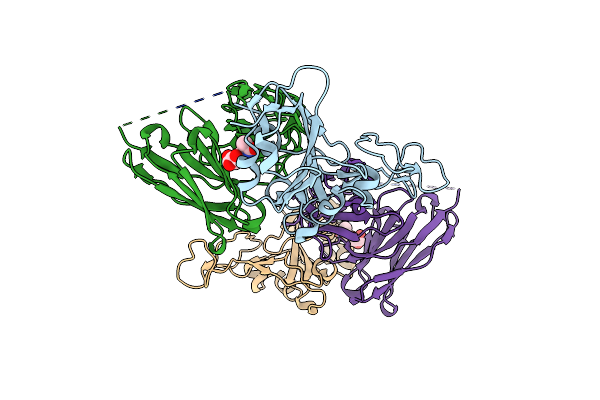
Crystal Structure Of Sars-Cov-2 Rbd In Complex With A Neutralizing Antibody Scfv
Organism: Homo sapiens, Severe acute respiratory syndrome coronavirus 2
Method: X-RAY DIFFRACTION Resolution:2.50 Å Release Date: 2021-03-31 Classification: VIRAL PROTEIN/ANTIVIRAL PROTEIN Ligands: CA, NAG |
 |
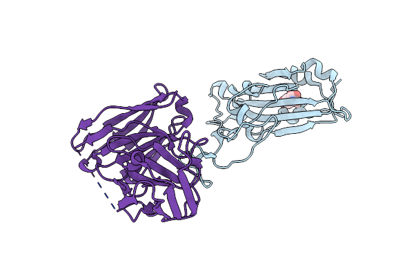
Crystal Structure Of Sars-Cov-2 Rbd In Complex With A Neutralizing Antibody Scfv
Organism: Severe acute respiratory syndrome coronavirus 2, Mus musculus
Method: X-RAY DIFFRACTION Resolution:2.20 Å Release Date: 2021-03-31 Classification: VIRAL PROTEIN/ANTIVIRAL PROTEIN Ligands: NAG |
 |
Crystal Structure Of Sars-Cov-2 Rbd In Complex With A Neutralizing Antibody Scfv
Organism: Severe acute respiratory syndrome coronavirus 2, Mus musculus
Method: X-RAY DIFFRACTION Resolution:2.10 Å Release Date: 2021-03-31 Classification: VIRAL PROTEIN/ANTIVIRAL PROTEIN Ligands: NAG |
 |
Crystal Structure Of The C-Terminal Periplasmic Domain Of Eceptc From Escherichia Coli- Complex With Zn
Organism: Escherichia coli
Method: X-RAY DIFFRACTION Resolution:2.10 Å Release Date: 2019-06-05 Classification: TRANSFERASE Ligands: ZN |
 |
Crystal Structure Of The C-Terminal Periplasmic Domain Of Eceptc From Escherichia Coli
Organism: Escherichia coli
Method: X-RAY DIFFRACTION Resolution:2.10 Å Release Date: 2018-12-26 Classification: TRANSFERASE |
 |
Crystal Structure Of The C-Terminal Periplasmic Domain Of Eceptc From Escherichia Coli Complex With Zn
Organism: Escherichia coli
Method: X-RAY DIFFRACTION Resolution:2.60 Å Release Date: 2018-12-26 Classification: TRANSFERASE Ligands: ZN, NA |