
Search Count: 95
 |
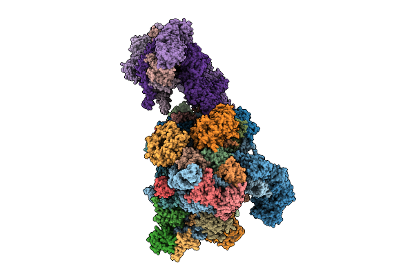
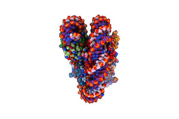
Organism: Oryctolagus cuniculus
Method: ELECTRON MICROSCOPY Release Date: 2025-12-10 Classification: TRANSLATION Ligands: SF4 |
 |
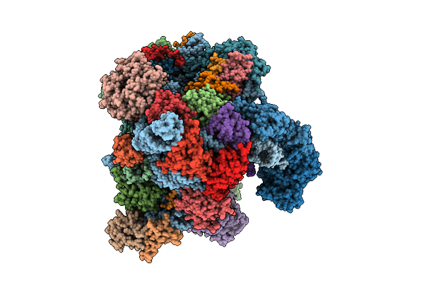
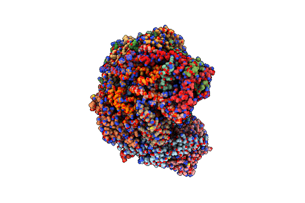
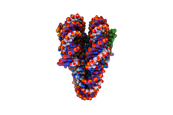
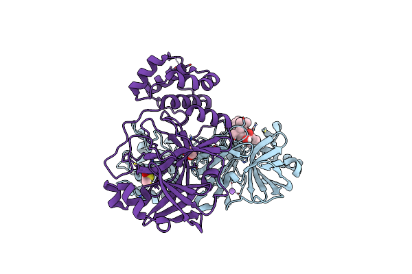
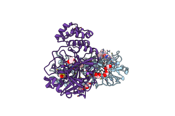
Late-Stage 48S Initiation Complex With Eif3 (Ls48S-Eif3 Ic) Guided By The Trans-Rna
Organism: Oryctolagus cuniculus
Method: ELECTRON MICROSCOPY Release Date: 2025-12-10 Classification: TRANSLATION Ligands: SF4 |
 |
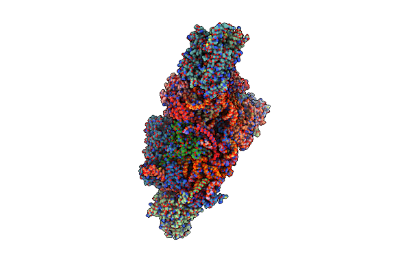
Organism: Brassica oleracea var. botrytis
Method: ELECTRON MICROSCOPY Release Date: 2025-10-15 Classification: RIBOSOME Ligands: MG, ATP, K, ZN |
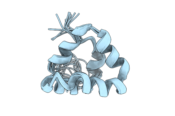 |
Organism: Homo sapiens
Method: SOLUTION NMR Release Date: 2025-10-01 Classification: METAL BINDING PROTEIN |
 |
Organism: Homo sapiens
Method: SOLUTION NMR Release Date: 2025-10-01 Classification: METAL BINDING PROTEIN |
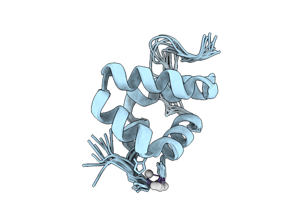 |
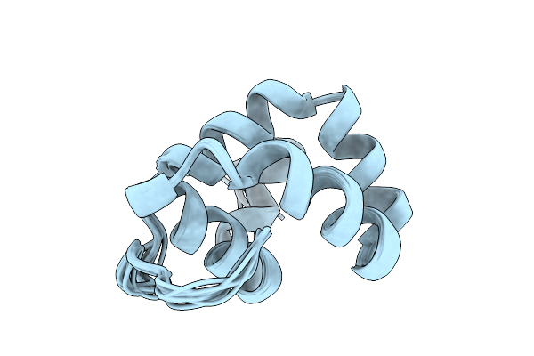
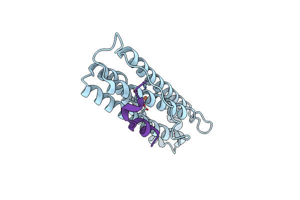
Solution Structure Of Silver Bound Xpc Binding Domain Of Hhr23B (Holo Form)
Organism: Homo sapiens
Method: SOLUTION NMR Release Date: 2025-10-01 Classification: METAL BINDING PROTEIN Ligands: AG |
 |
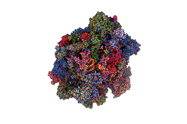
Organism: Brassica oleracea var. botrytis, Brassica oleracea var. oleracea, Arabidopsis thaliana
Method: ELECTRON MICROSCOPY Release Date: 2025-09-03 Classification: RIBOSOME Ligands: MG, ATP, CLM, K, ZN |
 |
Organism: Mus musculus, Influenza a virus h3n2
Method: X-RAY DIFFRACTION Release Date: 2025-02-05 Classification: IMMUNE SYSTEM Ligands: EDO, NAG |
 |
Organism: Mus musculus, Influenza a virus
Method: X-RAY DIFFRACTION Release Date: 2025-01-29 Classification: IMMUNE SYSTEM Ligands: EDO, NAG |
 |
Ssu(Head) Structure Derived From The Ssu Sample Of The Mitoribosome From T. Gondii.
Organism: Toxoplasma gondii
Method: ELECTRON MICROSCOPY Release Date: 2024-12-11 Classification: RIBOSOME |
 |
Ssu(Body) Structure Derived From The Ssu Sample Of The Mitoribosome From T. Gondii.
Organism: Toxoplasma gondii
Method: ELECTRON MICROSCOPY Release Date: 2024-12-11 Classification: RIBOSOME |
 |
Lsu Structure Derived From The Lsu Sample Of The Mitoribosome From T. Gondii.
Organism: Toxoplasma gondii
Method: ELECTRON MICROSCOPY Release Date: 2024-12-11 Classification: RIBOSOME |
 |
Organism: Homo sapiens, Synthetic construct
Method: ELECTRON MICROSCOPY Release Date: 2024-05-15 Classification: DNA BINDING PROTEIN/DNA |
 |
Organism: Homo sapiens, Synthetic construct
Method: ELECTRON MICROSCOPY Release Date: 2024-05-15 Classification: DNA BINDING PROTEIN/DNA |
 |
Organism: Homo sapiens, Synthetic construct
Method: ELECTRON MICROSCOPY Release Date: 2024-05-15 Classification: DNA BINDING PROTEIN/DNA |
 |
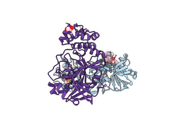
Structure Of The Ncoa4 (Nuclear Receptor Coactivator 4)-Fth1 (H-Ferritin) Complex
Organism: Homo sapiens
Method: ELECTRON MICROSCOPY Release Date: 2024-05-15 Classification: METAL TRANSPORT Ligands: FE |
 |
Crystal Structure Of The Main Protease (3Clpro/Mpro) Of Sars-Cov-2 Obtained In Presence Of 150 Micromolar Mg-132.
Organism: Severe acute respiratory syndrome coronavirus 2
Method: X-RAY DIFFRACTION Resolution:1.60 Å Release Date: 2024-05-01 Classification: VIRAL PROTEIN Ligands: ALD, CL, NA, DMS, EDO |
 |
Crystal Structure Of The Main Protease (3Clpro/Mpro) Of Sars-Cov-2 Obtained In Presence Of 75 Micromolar Mg-132.
Organism: Severe acute respiratory syndrome coronavirus 2
Method: X-RAY DIFFRACTION Resolution:1.85 Å Release Date: 2024-05-01 Classification: VIRAL PROTEIN |
 |
Crystal Structure Of The Main Protease (3Clpro/Mpro) Of Sars-Cov-2 Obtained In Presence Of 150 Micromolar X77.
Organism: Severe acute respiratory syndrome coronavirus 2
Method: X-RAY DIFFRACTION Resolution:1.63 Å Release Date: 2024-05-01 Classification: VIRAL PROTEIN Ligands: EDO, CL, X77, DMS, NA |
 |
Crystal Structure Of The Main Protease (3Clpro/Mpro) Of Sars-Cov-2 Obtained In Presence Of 75 Micromolar X77.
Organism: Severe acute respiratory syndrome coronavirus 2
Method: X-RAY DIFFRACTION Resolution:1.60 Å Release Date: 2024-05-01 Classification: VIRAL PROTEIN Ligands: CL, X77, EDO, DMS, NA |