
Search Count: 401
 |
Organism: Oryctolagus cuniculus
Method: ELECTRON MICROSCOPY Release Date: 2025-12-10 Classification: TRANSLATION Ligands: SF4 |
 |
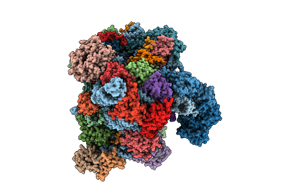
Late-Stage 48S Initiation Complex With Eif3 (Ls48S-Eif3 Ic) Guided By The Trans-Rna
Organism: Oryctolagus cuniculus
Method: ELECTRON MICROSCOPY Release Date: 2025-12-10 Classification: TRANSLATION Ligands: SF4 |
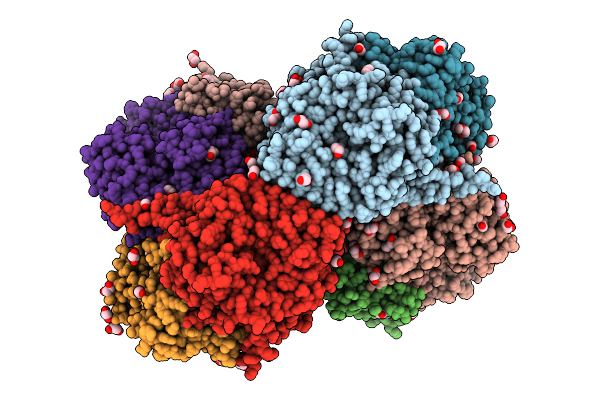 |
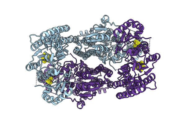
Organism: Azotobacter vinelandii dj
Method: X-RAY DIFFRACTION Release Date: 2025-12-03 Classification: METAL BINDING PROTEIN Ligands: SF4, S5Q, PEG, EDO, 1PE, PGE, MG, PG4 |
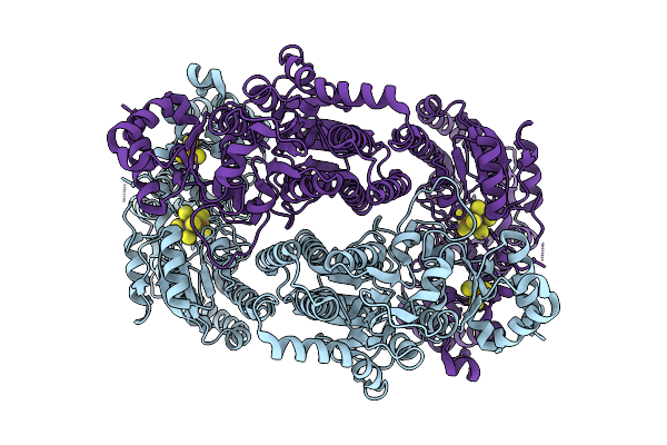 |
Organism: Geobacter metallireducens
Method: ELECTRON MICROSCOPY Release Date: 2025-11-05 Classification: METAL BINDING PROTEIN Ligands: SF4, S5Q |
 |
Organism: Geobacter metallireducens
Method: ELECTRON MICROSCOPY Release Date: 2025-11-05 Classification: METAL BINDING PROTEIN Ligands: SF4, S5Q |
 |
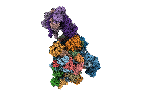
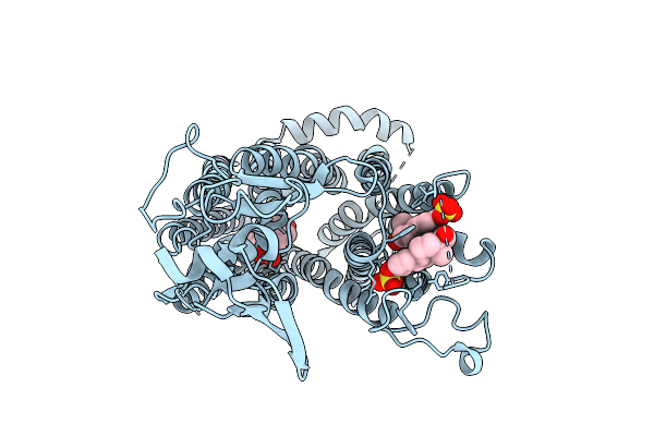
Organism: Brassica oleracea var. botrytis
Method: ELECTRON MICROSCOPY Release Date: 2025-10-15 Classification: RIBOSOME Ligands: MG, ATP, K, ZN |
 |
Crystal Structure Of The A/Viet Nam/1203/2004(H5N1) Influenza Virus Hemagglutinin In Complex With Cyclic Peptide Iha-100
Organism: Influenza a virus, Homo sapiens
Method: X-RAY DIFFRACTION Release Date: 2025-10-08 Classification: VIRAL PROTEIN |
 |
Organism: Homo sapiens
Method: SOLUTION NMR Release Date: 2025-10-01 Classification: METAL BINDING PROTEIN |
 |
Organism: Homo sapiens
Method: SOLUTION NMR Release Date: 2025-10-01 Classification: METAL BINDING PROTEIN |
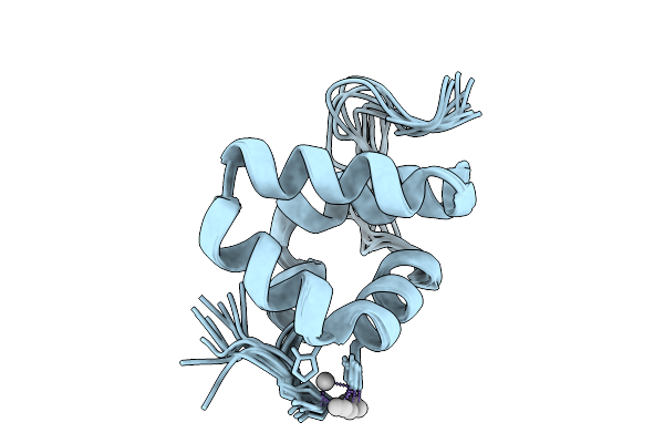 |
Solution Structure Of Silver Bound Xpc Binding Domain Of Hhr23B (Holo Form)
Organism: Homo sapiens
Method: SOLUTION NMR Release Date: 2025-10-01 Classification: METAL BINDING PROTEIN Ligands: AG |
 |
Structure Of A De Novo Designed Interleukin-21 Mimetic Complex With Il-21R And Il-2Rg
Organism: Synthetic construct, Homo sapiens
Method: X-RAY DIFFRACTION Release Date: 2025-09-24 Classification: DE NOVO PROTEIN/IMMUNE SYSTEM Ligands: NAG, EDO |
 |
Organism: Brassica oleracea var. botrytis, Brassica oleracea var. oleracea, Arabidopsis thaliana
Method: ELECTRON MICROSCOPY Release Date: 2025-09-03 Classification: RIBOSOME Ligands: MG, ATP, CLM, K, ZN |
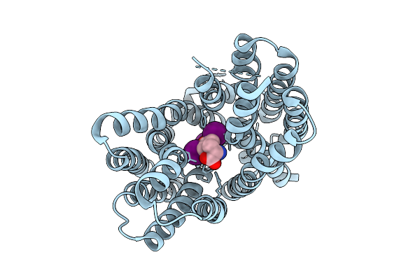 |
Organism: Homo sapiens
Method: ELECTRON MICROSCOPY Release Date: 2025-07-16 Classification: TRANSPORT PROTEIN Ligands: T3 |
 |
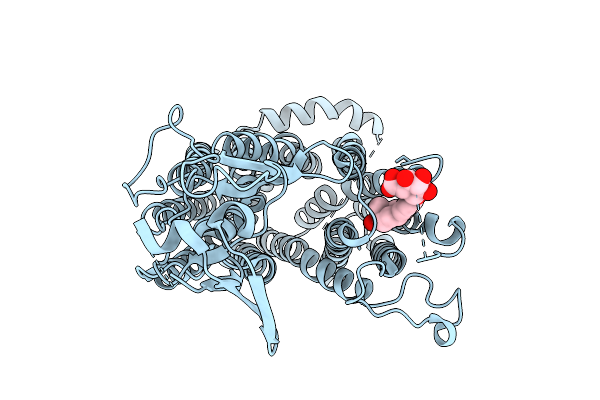
Cryo-Em Structure Of Human Oatp1C1 F240A Mutant In Complex With Estrone 3-Sulfate
Organism: Homo sapiens
Method: ELECTRON MICROSCOPY Release Date: 2025-07-16 Classification: TRANSPORT PROTEIN Ligands: FY5 |
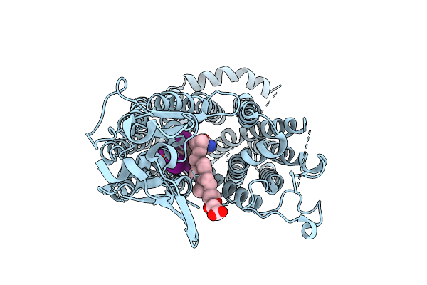 |
Organism: Homo sapiens
Method: ELECTRON MICROSCOPY Release Date: 2025-07-16 Classification: TRANSPORT PROTEIN Ligands: T44, Y01 |
 |
Cryo-Em Structure Of Human Oatp1C1 F240A Mutant In Complex With Estrone 3-Glucuronide
Organism: Homo sapiens
Method: ELECTRON MICROSCOPY Release Date: 2025-07-16 Classification: TRANSPORT PROTEIN Ligands: E3G |
 |
 |
 |
 |
Organism: Homo sapiens
Method: ELECTRON MICROSCOPY Release Date: 2025-07-16 Classification: RNA BINDING PROTEIN |