
Search Count: 115
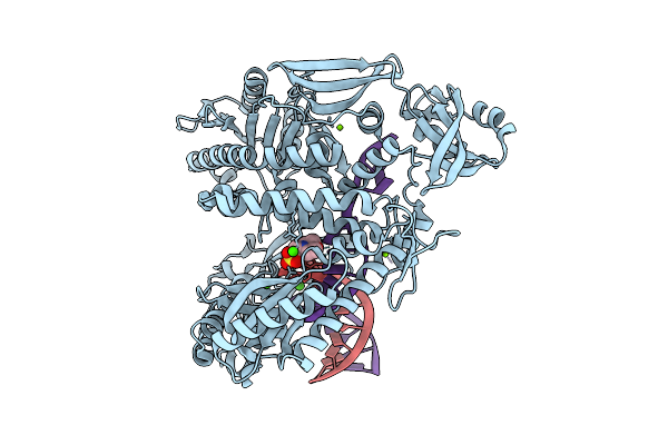 |
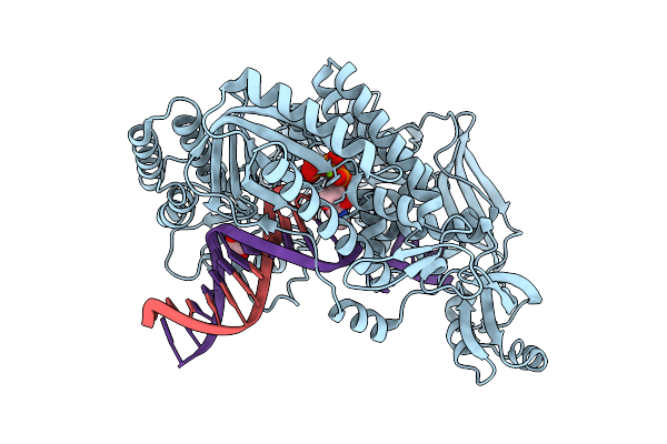
Organism: Thermococcus kodakarensis, Synthetic construct
Method: X-RAY DIFFRACTION Release Date: 2025-12-10 Classification: TRANSFERASE/DNA Ligands: CA, MG, MES |
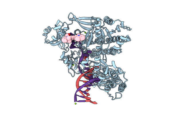 |
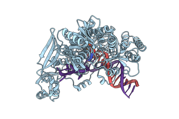
Organism: Thermococcus kodakarensis, Synthetic construct
Method: X-RAY DIFFRACTION Release Date: 2025-12-10 Classification: TRANSFERASE/DNA Ligands: MG, 5CY, GOL |
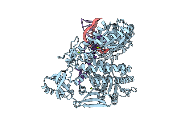 |
Organism: Thermococcus kodakarensis, Synthetic construct
Method: X-RAY DIFFRACTION Release Date: 2025-12-10 Classification: TRANSFERASE/DNA Ligands: MG |
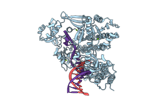 |
Organism: Thermococcus kodakarensis, Synthetic construct
Method: X-RAY DIFFRACTION Release Date: 2025-12-10 Classification: TRANSFERASE/DNA Ligands: MG |
 |
Organism: Thermococcus kodakarensis, Synthetic construct
Method: X-RAY DIFFRACTION Release Date: 2025-12-10 Classification: TRANSFERASE/DNA Ligands: 9O7, MG, IOD, EDO |
 |
Organism: Thermococcus kodakarensis, Synthetic construct
Method: X-RAY DIFFRACTION Release Date: 2025-12-10 Classification: TRANSFERASE/DNA Ligands: 9O7, MG, IOD, CL |
 |
Organism: Homo sapiens
Method: ELECTRON MICROSCOPY Release Date: 2025-07-09 Classification: MOTOR PROTEIN Ligands: ADP, ATP, MG |
 |
Organism: Homo sapiens
Method: ELECTRON MICROSCOPY Release Date: 2025-07-09 Classification: MOTOR PROTEIN Ligands: ADP, MG, ATP |
 |
Cryo-Em Structure Of Human Cytoplasmic Dynein-1 Bound To Lis1 In The Presence Of Atp
Organism: Homo sapiens
Method: ELECTRON MICROSCOPY Release Date: 2025-07-09 Classification: MOTOR PROTEIN Ligands: ADP, MG, ATP |
 |
Organism: Homo sapiens
Method: ELECTRON MICROSCOPY Release Date: 2025-07-09 Classification: MOTOR PROTEIN Ligands: ADP, ATP, MG |
 |
Cryo-Em Structure Of Human Cytoplasmic Dynein-1 Bound To Lis1 In The Presence Of Atp
Organism: Homo sapiens
Method: ELECTRON MICROSCOPY Release Date: 2025-07-09 Classification: MOTOR PROTEIN Ligands: ADP, ATP, MG |
 |
Organism: Homo sapiens
Method: ELECTRON MICROSCOPY Release Date: 2025-07-09 Classification: MOTOR PROTEIN Ligands: ADP, ATP, MG |
 |
Organism: Homo sapiens
Method: ELECTRON MICROSCOPY Release Date: 2025-07-09 Classification: MOTOR PROTEIN Ligands: ADP, ATP |
 |
Cryo-Em Structure Of Human Cytoplasmic Dynein-1 Bound To Lis1 In The Presence Of Atp
Organism: Homo sapiens
Method: ELECTRON MICROSCOPY Release Date: 2025-07-09 Classification: MOTOR PROTEIN Ligands: ADP, ATP, MG |
 |
Organism: Homo sapiens
Method: ELECTRON MICROSCOPY Release Date: 2025-07-09 Classification: MOTOR PROTEIN |
 |
Organism: Homo sapiens
Method: ELECTRON MICROSCOPY Release Date: 2025-07-09 Classification: MOTOR PROTEIN |
 |
Cryoem Structures Of Yeast Cytoplasmic Dynein In The Presence Of Atp And Lis1.
Organism: Saccharomyces cerevisiae
Method: ELECTRON MICROSCOPY Release Date: 2025-06-04 Classification: MOTOR PROTEIN Ligands: ATP, ADP |
 |
Cryoem Structures Of Yeast Cytoplasmic Dynein In The Presence Of Atp And Lis1.
Organism: Saccharomyces cerevisiae
Method: ELECTRON MICROSCOPY Release Date: 2025-06-04 Classification: MOTOR PROTEIN Ligands: ATP, ADP |
 |
Cryoem Structures Of Yeast Cytoplasmic Dynein In The Presence Of Atp And Lis1.
Organism: Saccharomyces cerevisiae
Method: ELECTRON MICROSCOPY Release Date: 2025-06-04 Classification: MOTOR PROTEIN Ligands: ATP, ADP |
 |
Cryoem Structures Of Yeast Cytoplasmic Dynein In The Presence Of Atp And Lis1.
Organism: Saccharomyces cerevisiae
Method: ELECTRON MICROSCOPY Release Date: 2025-06-04 Classification: MOTOR PROTEIN Ligands: ATP, ADP, MG |