
Search Count: 32
 |
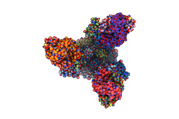
Organism: Merbecovirus, Pteronotus davyi
Method: ELECTRON MICROSCOPY Release Date: 2025-03-05 Classification: VIRAL PROTEIN Ligands: NAG |
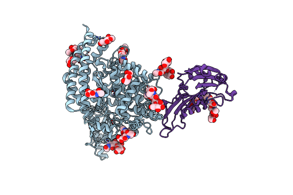 |
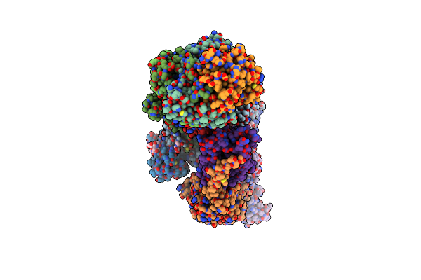
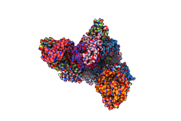
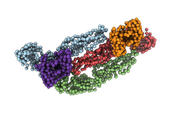
Merbecovirus Pnnl2018B Spike Glycoprotein Rbd Bound To The P. Nathusii Ace2
Organism: Pipistrellus nathusii, Merbecovirus
Method: ELECTRON MICROSCOPY Release Date: 2025-02-19 Classification: VIRAL PROTEIN Ligands: NAG, ZN |
 |
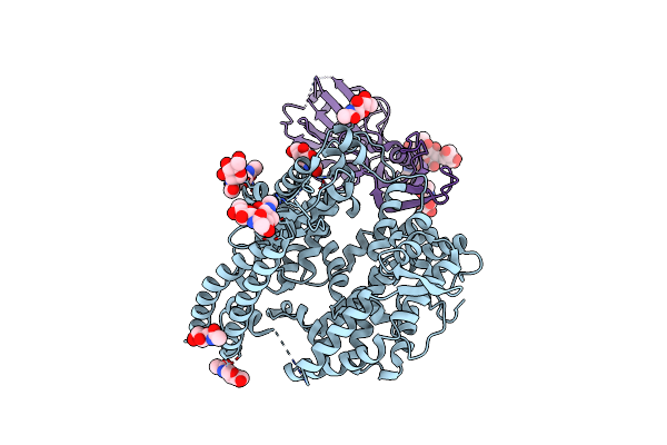
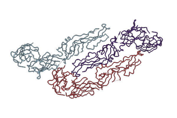
Organism: Pipistrellus nathusii, Middle east respiratory syndrome-related coronavirus
Method: ELECTRON MICROSCOPY Release Date: 2025-02-12 Classification: VIRAL PROTEIN/HYDROLASE Ligands: NAG, ZN |
 |
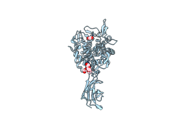
Crystal Structure Of Hiv-1 Capsid N-Terminal Domain In The Presence Of Lenacapavir
Organism: Human immunodeficiency virus 1
Method: X-RAY DIFFRACTION Resolution:2.00 Å Release Date: 2025-01-08 Classification: VIRAL PROTEIN |
 |
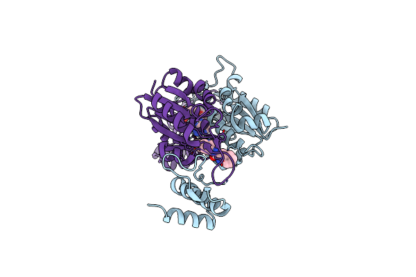
Organism: Severe acute respiratory syndrome coronavirus 2, Homo sapiens
Method: ELECTRON MICROSCOPY Release Date: 2024-11-20 Classification: VIRAL PROTEIN Ligands: NAG |
 |
Sars-Cov-2 S + S2L20 (Local Refinement Of Ntd And S2L20 Fab Variable Region)
Organism: Homo sapiens, Severe acute respiratory syndrome coronavirus 2
Method: ELECTRON MICROSCOPY Release Date: 2024-11-20 Classification: VIRAL PROTEIN/IMMUNE SYSTEM Ligands: NAG |
 |
Organism: Homo sapiens
Method: ELECTRON MICROSCOPY Release Date: 2024-08-21 Classification: MEMBRANE PROTEIN Ligands: NAG |
 |
Organism: Homo sapiens
Method: ELECTRON MICROSCOPY Release Date: 2024-08-21 Classification: MEMBRANE PROTEIN Ligands: NAG |
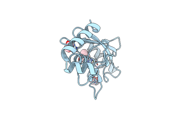 |
Identification Of Small-Molecule Binding Sites Of A Ubiquitin-Conjugating Enzyme-Ube2T Through Fragment-Based Screening
Organism: Homo sapiens
Method: X-RAY DIFFRACTION Resolution:1.54 Å Release Date: 2024-02-28 Classification: LIGASE Ligands: V23, EDO |
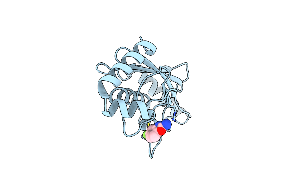 |
Identification Of Small-Molecule Binding Sites Of A Ubiquitin-Conjugating Enzyme-Ube2T Through Fragment-Based Screening
Organism: Homo sapiens
Method: X-RAY DIFFRACTION Resolution:1.70 Å Release Date: 2024-02-28 Classification: LIGASE Ligands: V2R |
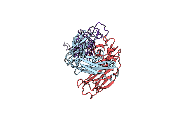 |
Sars-Cov-2 Spike In Complex With The Zb8 Neutralizing Antibody Fab (Focused Refinement On Fab-Rbd)
Organism: Homo sapiens, Severe acute respiratory syndrome coronavirus 2
Method: ELECTRON MICROSCOPY Release Date: 2023-01-18 Classification: IMMUNE SYSTEM/VIRAL PROTEIN |
 |
Organism: Severe acute respiratory syndrome coronavirus 2, Homo sapiens
Method: ELECTRON MICROSCOPY Release Date: 2023-01-18 Classification: VIRAL PROTEIN/IMMUNE SYSTEM Ligands: NAG |
 |
Organism: Severe acute respiratory syndrome coronavirus 2, Homo sapiens
Method: ELECTRON MICROSCOPY Release Date: 2023-01-18 Classification: VIRAL PROTEIN/IMMUNE SYSTEM Ligands: NAG |
 |
Organism: Mus musculus
Method: SOLUTION NMR Release Date: 2020-09-09 Classification: ANTIVIRAL PROTEIN |
 |
Crystal Structure Of A Family 76 Glycoside Hydrolase From A Bovine Bacteroides Thetaiotaomicron Strain
Organism: Bacteroides thetaiotaomicron
Method: X-RAY DIFFRACTION Resolution:1.40 Å Release Date: 2020-04-29 Classification: HYDROLASE Ligands: P6G, EDO |
 |
Crystal Structure Of Trmd From Pseudomonas Aeruginosa In Complex With Active-Site Inhibitor
Organism: Pseudomonas aeruginosa ucbpp-pa14
Method: X-RAY DIFFRACTION Resolution:2.75 Å Release Date: 2019-08-14 Classification: TRANSFERASE Ligands: SAM, 9WO |
 |
Organism: Enterovirus d68
Method: X-RAY DIFFRACTION Resolution:2.15 Å Release Date: 2019-04-17 Classification: RNA BINDING PROTEIN Ligands: GOL, PEG |
 |
Organism: Homo sapiens
Method: X-RAY DIFFRACTION Resolution:2.50 Å Release Date: 2019-01-23 Classification: CELL ADHESION |
 |
Three-Dimensional Structure Of Dengue Virus Serotype 1 Complexed With 2 Hmab 14C10 Fab
Organism: Dengue virus 1, Homo sapiens
Method: ELECTRON MICROSCOPY Resolution:7.00 Å Release Date: 2013-10-16 Classification: VIRUS |
 |
Three-Dimensional Structure Of Dengue Virus Serotype 1 Complexed With Hmab 14C10 Fab
|