
Search Count: 131
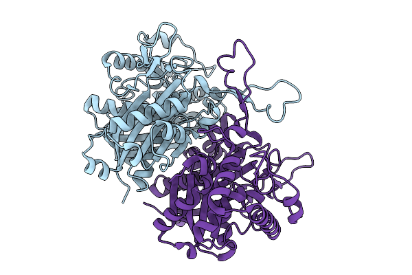 |
Organism: Burkholderia sp. rf2-non_bp3
Method: X-RAY DIFFRACTION Release Date: 2025-08-27 Classification: LYASE |
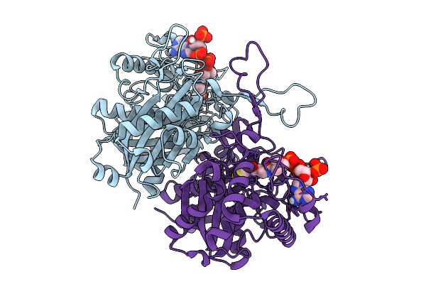 |
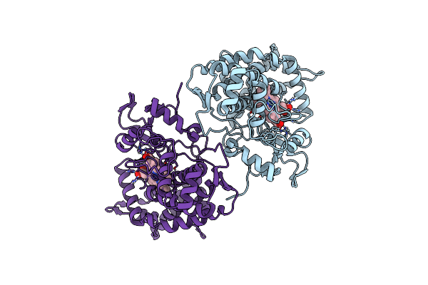
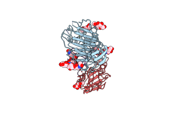
Crystal Structure Of Polyketoacyl-Coa Thiolase From Burkholderia Sp In Complex With Butyryl-Coa
Organism: Burkholderia sp. rf2-non_bp3
Method: X-RAY DIFFRACTION Release Date: 2025-08-27 Classification: LYASE Ligands: COA |
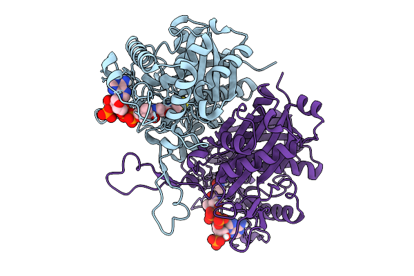 |
Crystal Structure Of Polyketoacyl-Coa Thiolase From Burkholderia Sp. In Complex With Acetyl-Coa
Organism: Burkholderia sp. rf2-non_bp3
Method: X-RAY DIFFRACTION Release Date: 2025-08-27 Classification: LYASE Ligands: COA |
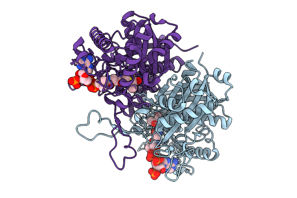 |
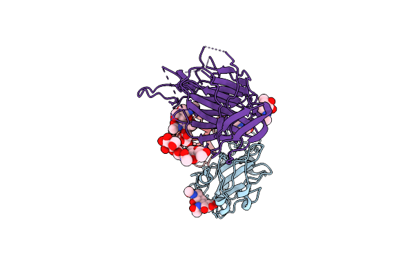
Crystal Structure Of Polyketoacyl-Coa Thiolase From Burkholderia Sp. In Complex With Acetoacetyl-Coa
Organism: Burkholderia sp. rf2-non_bp3
Method: X-RAY DIFFRACTION Release Date: 2025-08-27 Classification: LYASE Ligands: COA, A1BNQ |
 |
Organism: Severe acute respiratory syndrome coronavirus 2, Enterobacteria phage t4, Synthetic construct, Homo sapiens
Method: ELECTRON MICROSCOPY Release Date: 2025-01-22 Classification: VIRAL PROTEIN |
 |
Organism: Severe acute respiratory syndrome coronavirus 2, Enterobacteria phage t4, Synthetic construct, Homo sapiens
Method: ELECTRON MICROSCOPY Release Date: 2025-01-22 Classification: VIRAL PROTEIN |
 |
Organism: Severe acute respiratory syndrome coronavirus 2, Enterobacteria phage t4, Synthetic construct, Homo sapiens
Method: ELECTRON MICROSCOPY Release Date: 2025-01-22 Classification: VIRAL PROTEIN |
 |
Organism: Severe acute respiratory syndrome coronavirus 2, Tequatrovirus t4, Synthetic construct, Homo sapiens
Method: ELECTRON MICROSCOPY Release Date: 2025-01-22 Classification: VIRAL PROTEIN |
 |
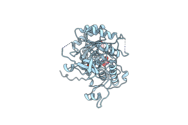
Crystal Structure Of Polyketide Synthase (Pks) Thioreductase Domain From Streptomyces Coelicolor
Organism: Streptomyces coelicolor
Method: X-RAY DIFFRACTION Release Date: 2024-11-27 Classification: BIOSYNTHETIC PROTEIN Ligands: NDP, GOL, BTB |
 |
Organism: Lama glama, Bos taurus
Method: X-RAY DIFFRACTION Resolution:3.70 Å Release Date: 2023-08-30 Classification: MEMBRANE PROTEIN Ligands: RET |
 |
Organism: Lama glama, Bos taurus
Method: X-RAY DIFFRACTION Resolution:3.71 Å Release Date: 2023-08-30 Classification: MEMBRANE PROTEIN |
 |
Organism: Lama glama, Bos taurus
Method: X-RAY DIFFRACTION Resolution:4.25 Å Release Date: 2023-08-30 Classification: MEMBRANE PROTEIN |
 |
Organism: Metagenome
Method: X-RAY DIFFRACTION Resolution:1.53 Å Release Date: 2023-05-10 Classification: BIOSYNTHETIC PROTEIN Ligands: HEM |
 |
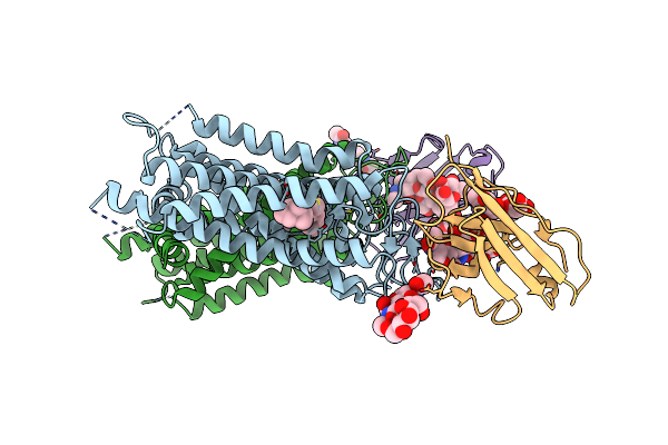
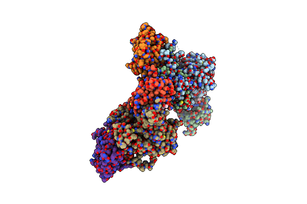
Cryo-Em Structure Of Sars-Cov-2 Spike (Hexapro Variant) In Complex With Nanobody W25 (Map 3, Focus Refinement On Rbd, W25 And Adjacent Ntd)
Organism: Severe acute respiratory syndrome coronavirus 2, Vicugna pacos
Method: ELECTRON MICROSCOPY Release Date: 2023-03-08 Classification: VIRAL PROTEIN Ligands: NAG |
 |
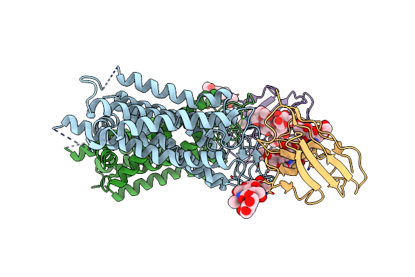
Cryo-Em Structure Of Sars-Cov-2 Spike (Omicron Ba.1 Variant) In Complex With Nanobody W25 (Map 5, Focus Refinement On Rbd, W25 And Adjacent Ntd)
Organism: Severe acute respiratory syndrome coronavirus 2, Vicugna pacos
Method: ELECTRON MICROSCOPY Release Date: 2023-03-08 Classification: VIRAL PROTEIN Ligands: NAG |
 |
Organism: Bacillus subtilis
Method: X-RAY DIFFRACTION Resolution:2.44 Å Release Date: 2022-09-14 Classification: SIGNALING PROTEIN Ligands: B4P, MG |
 |
Organism: Bacillus subtilis (strain 168)
Method: X-RAY DIFFRACTION Resolution:1.76 Å Release Date: 2022-09-14 Classification: SIGNALING PROTEIN Ligands: PO4, GOL |
 |
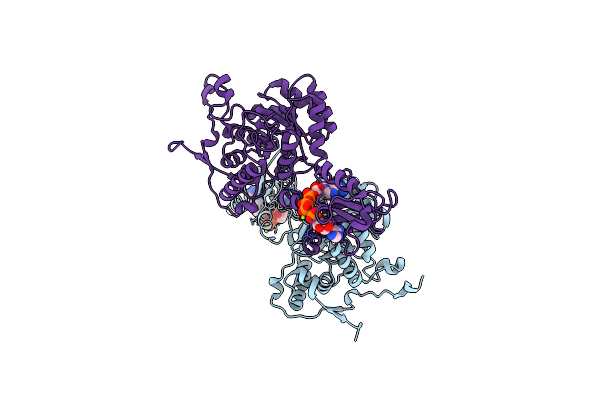
Structure Of Full Length Human Ampk (A2B1G1) In Complex With A Small Molecule Activator Msg011
Organism: Homo sapiens
Method: X-RAY DIFFRACTION Resolution:2.95 Å Release Date: 2022-06-29 Classification: SIGNALING PROTEIN Ligands: 4O7, ZQV, AMP |
 |
Organism: Bacillus subtilis
Method: X-RAY DIFFRACTION Resolution:2.45 Å Release Date: 2021-12-22 Classification: DNA BINDING PROTEIN Ligands: G4P |
 |
Organism: Homo sapiens
Method: X-RAY DIFFRACTION Resolution:2.24 Å Release Date: 2021-09-29 Classification: TRANSFERASE Ligands: 7QB, PEG, EDO, CA |