
Search Count: 24
 |
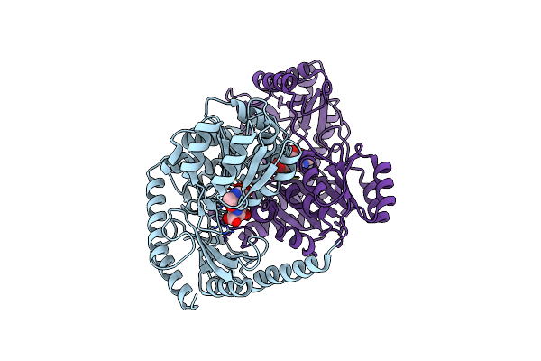
Human Cx36/Gjd2 Gap Junction Channel With Pore-Lining N-Terminal Helices In Porcine Brain Lipids.
Organism: Homo sapiens
Method: ELECTRON MICROSCOPY Release Date: 2024-11-06 Classification: MEMBRANE PROTEIN Ligands: MC3, Y01 |
 |
Organism: Homo sapiens
Method: ELECTRON MICROSCOPY Release Date: 2024-11-06 Classification: MEMBRANE PROTEIN Ligands: MC3 |
 |
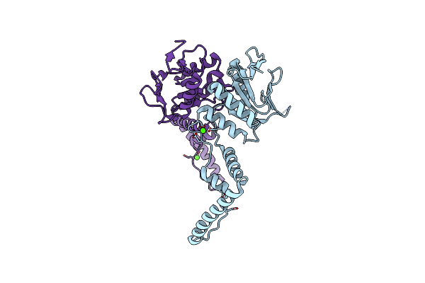
Organism: Homo sapiens
Method: ELECTRON MICROSCOPY Release Date: 2024-11-06 Classification: MEMBRANE PROTEIN Ligands: MC3, ACD |
 |
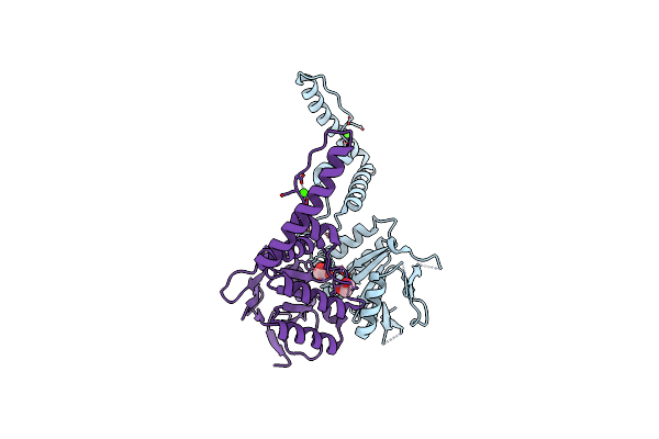
Organism: Homo sapiens
Method: ELECTRON MICROSCOPY Release Date: 2024-11-06 Classification: MEMBRANE PROTEIN Ligands: MC3, HE2 |
 |
Organism: Homo sapiens
Method: ELECTRON MICROSCOPY Release Date: 2024-11-06 Classification: MEMBRANE PROTEIN Ligands: YMZ, MC3 |
 |
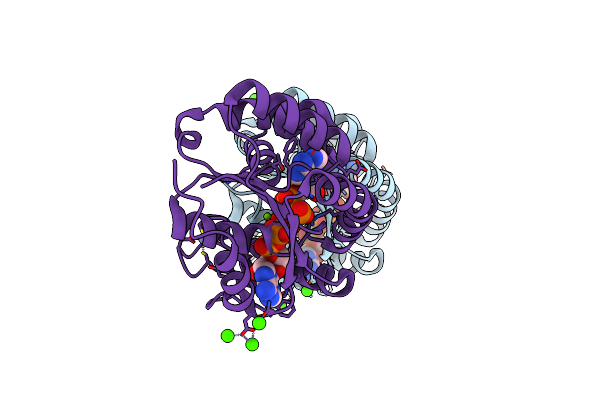
Human Cx36/Gjd2 (Ala14-Deleted Mutant) Gap Junction Channel In Porcine Brain Lipids
Organism: Homo sapiens
Method: ELECTRON MICROSCOPY Release Date: 2024-11-06 Classification: MEMBRANE PROTEIN Ligands: MC3 |
 |
Human Cx36/Gjd2 (Ala14 Deletion Mutant) Gap Junction Channel Prepared With Mefloquine, Showing No Bound Mefloquine
Organism: Homo sapiens
Method: ELECTRON MICROSCOPY Release Date: 2024-11-06 Classification: MEMBRANE PROTEIN Ligands: MC3 |
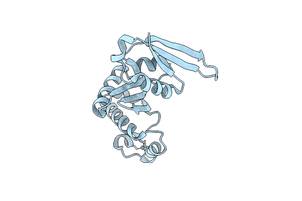 |
Organism: Saccharolobus solfataricus p2
Method: X-RAY DIFFRACTION Resolution:1.68 Å Release Date: 2024-10-02 Classification: RNA BINDING PROTEIN Ligands: ZN |
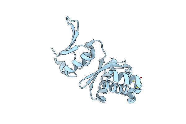 |
Organism: Sulfolobus acidocaldarius dsm 639
Method: X-RAY DIFFRACTION Resolution:3.10 Å Release Date: 2024-09-11 Classification: UNKNOWN FUNCTION Ligands: ZN |
 |
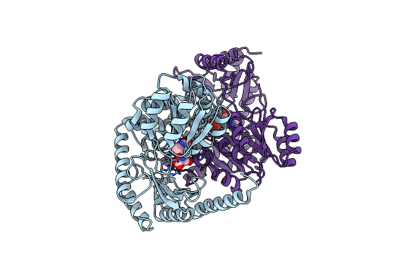
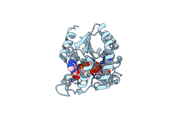
Organism: Streptomyces albogriseolus 1-36
Method: X-RAY DIFFRACTION Resolution:2.49 Å Release Date: 2024-05-01 Classification: TRANSFERASE Ligands: PLP |
 |
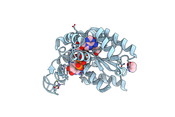
Crystal Structure Of Alpha-Oxoamine Synthase Alb29 With Plp Cofactor And L-Glutamate
Organism: Streptomyces albogriseolus 1-36
Method: X-RAY DIFFRACTION Resolution:2.27 Å Release Date: 2024-05-01 Classification: TRANSFERASE Ligands: PGU, PLP |
 |
Crystal Structure Of Alpha-Oxoamine Synthase Alb29 With Plp Cofactor And L-Glutamate
Organism: Streptomyces albogriseolus 1-36
Method: X-RAY DIFFRACTION Resolution:2.70 Å Release Date: 2024-05-01 Classification: TRANSFERASE Ligands: PGU, PLP |
 |
Organism: Streptomyces albogriseolus 1-36
Method: X-RAY DIFFRACTION Resolution:2.79 Å Release Date: 2024-05-01 Classification: TRANSFERASE Ligands: PLP, PLR |
 |
Organism: Francisella tularensis subsp. novicida (strain u112)
Method: X-RAY DIFFRACTION Resolution:2.49 Å Release Date: 2017-08-16 Classification: TRANSCRIPTION |
 |
Organism: Bacillus subtilis
Method: X-RAY DIFFRACTION Resolution:1.70 Å Release Date: 2014-03-26 Classification: TRANSFERASE Ligands: ZN, MG, AP5, CA |
 |
Organism: Bacillus subtilis
Method: X-RAY DIFFRACTION Resolution:1.45 Å Release Date: 2014-03-26 Classification: TRANSFERASE Ligands: ZN, MG, AP5 |
 |
Organism: Bacillus subtilis
Method: X-RAY DIFFRACTION Resolution:1.50 Å Release Date: 2014-03-26 Classification: TRANSFERASE Ligands: ZN, AP5, EDO |
 |
Organism: Streptococcus pyogenes
Method: X-RAY DIFFRACTION Resolution:2.90 Å Release Date: 2012-06-27 Classification: DNA BINDING PROTEIN Ligands: CA |
 |
Organism: Streptococcus pyogenes
Method: X-RAY DIFFRACTION Resolution:2.20 Å Release Date: 2012-05-30 Classification: DNA BINDING PROTEIN Ligands: CA, EDO |
 |