
Search Count: 66
 |
Organism: Arabidopsis thaliana
Method: X-RAY DIFFRACTION Release Date: 2025-08-13 Classification: PROTEIN TRANSPORT Ligands: MAN, NAG |
 |
Organism: Haemophilus influenzae (strain 86-028np), Escherichia coli
Method: X-RAY DIFFRACTION Resolution:3.15 Å Release Date: 2021-10-27 Classification: PEPTIDE BINDING PROTEIN Ligands: HEM |
 |
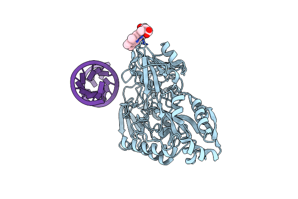
Nontypeable Haemophillus Influenzae Sapa In Complex With Double Stranded Rna
Organism: Haemophilus influenzae (strain 86-028np), Escherichia coli
Method: X-RAY DIFFRACTION Resolution:2.62 Å Release Date: 2021-10-27 Classification: PEPTIDE BINDING PROTEIN |
 |
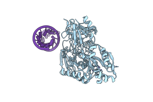
Nontypeable Haemophillus Influenzae Sapa In Open And Closed Conformations, In Complex With Double Stranded Rna
Organism: Haemophilus influenzae (strain 86-028np), Escherichia coli
Method: X-RAY DIFFRACTION Resolution:2.61 Å Release Date: 2021-10-27 Classification: PEPTIDE BINDING PROTEIN Ligands: CA |
 |
Organism: Homo sapiens
Method: X-RAY DIFFRACTION Resolution:2.10 Å Release Date: 2020-11-25 Classification: TRANSFERASE |
 |
Organism: Bos taurus
Method: X-RAY DIFFRACTION Resolution:2.15 Å Release Date: 2019-05-29 Classification: IMMUNE SYSTEM |
 |
Organism: Bos taurus
Method: X-RAY DIFFRACTION Resolution:2.12 Å Release Date: 2019-05-29 Classification: IMMUNE SYSTEM Ligands: CL |
 |
Organism: Bos taurus
Method: X-RAY DIFFRACTION Resolution:1.89 Å Release Date: 2019-05-29 Classification: IMMUNE SYSTEM Ligands: GOL, SO4 |
 |
Organism: Bos taurus
Method: X-RAY DIFFRACTION Resolution:2.62 Å Release Date: 2019-05-29 Classification: IMMUNE SYSTEM Ligands: GOL |
 |
Organism: Plasmodium vivax
Method: X-RAY DIFFRACTION Resolution:2.90 Å Release Date: 2018-08-29 Classification: TRANSFERASE Ligands: HH2, APC, PAB, MG, PE0, 5AD |
 |
Organism: Homo sapiens
Method: X-RAY DIFFRACTION Resolution:2.05 Å Release Date: 2018-03-07 Classification: TRANSCRIPTION |
 |
Cocrystal Structure Of Fc Gamma Receptor Iiia Interacting With Affimer F4, A Specific Binding Protein Which Blocks Igg Binding To The Receptor.
Organism: Homo sapiens, Synthetic construct
Method: X-RAY DIFFRACTION Resolution:2.35 Å Release Date: 2017-12-13 Classification: IMMUNE SYSTEM Ligands: NAG, SO4, PEG, CL |
 |
Cocrystal Structure Of Fc Gamma Receptor Iiia Interacting With Affimer G3, A Specific Binding Protein Which Blocks Igg Binding To The Receptor.
Organism: Homo sapiens, Synthetic construct
Method: X-RAY DIFFRACTION Resolution:2.35 Å Release Date: 2017-12-13 Classification: IMMUNE SYSTEM Ligands: NAG, PEG, GOL, PG4 |
 |
Crystal Structure Of Isoform 2 Of Purine Nucleoside Phosphorylase Complexed With Adenine
Organism: Schistosoma mansoni
Method: X-RAY DIFFRACTION Resolution:1.90 Å Release Date: 2017-10-11 Classification: TRANSFERASE Ligands: ADE, DMS |
 |
Crystal Structure Of Isoform 2 Of Purine Nucleoside Phosphorylase Complexed With Cytidine
Organism: Schistosoma mansoni
Method: X-RAY DIFFRACTION Resolution:2.10 Å Release Date: 2017-10-11 Classification: TRANSFERASE Ligands: CTN, SO4 |
 |
Crystal Structure Of Isoform 2 Of Purine Nucleoside Phosphorylase Complexed With Hypoxanthine
Organism: Schistosoma mansoni
Method: X-RAY DIFFRACTION Resolution:2.10 Å Release Date: 2017-10-11 Classification: TRANSFERASE Ligands: HPA, DMS |
 |
Crystal Structure Of Nucleoside Diphosphate Kinase From Schistosoma Mansoni Is Space Group P6322
Organism: Schistosoma mansoni
Method: X-RAY DIFFRACTION Resolution:1.90 Å Release Date: 2017-08-09 Classification: TRANSFERASE |
 |
Crystal Structure Of Isoform 2 Of Purine Nucleoside Phosphorylase From Schistosoma Mansoni In Complex With Cytosine
Organism: Schistosoma mansoni
Method: X-RAY DIFFRACTION Resolution:1.36 Å Release Date: 2017-08-09 Classification: TRANSFERASE Ligands: CYT, EDO |
 |
Crystal Structure Of Isoform 2 Of Purine Nucleoside Phosphorylase From Schistosoma Mansoni In Complex With Cytosine And Ribose-1-Phosphate
Organism: Schistosoma mansoni
Method: X-RAY DIFFRACTION Resolution:1.42 Å Release Date: 2017-08-09 Classification: TRANSFERASE Ligands: CYT, R1P |
 |
Crystal Structure Of Nucleoside Diphosphate Kinase From Schistosoma Mansoni In Complex With Adp
Organism: Schistosoma mansoni
Method: X-RAY DIFFRACTION Resolution:2.11 Å Release Date: 2017-06-21 Classification: TRANSFERASE Ligands: ADP |