
Search Count: 40
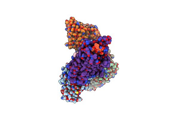 |
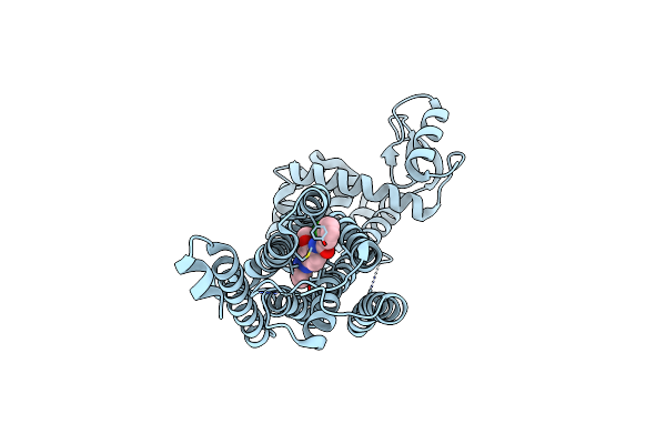
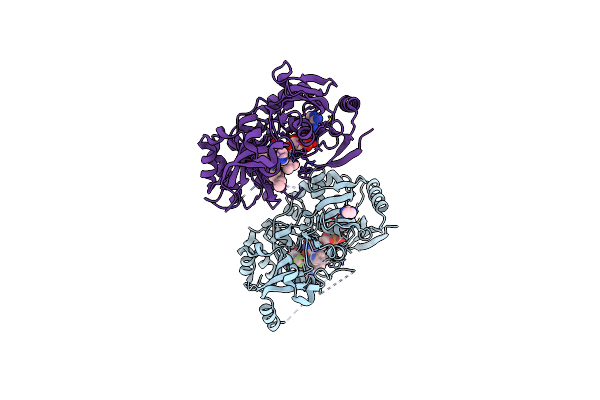
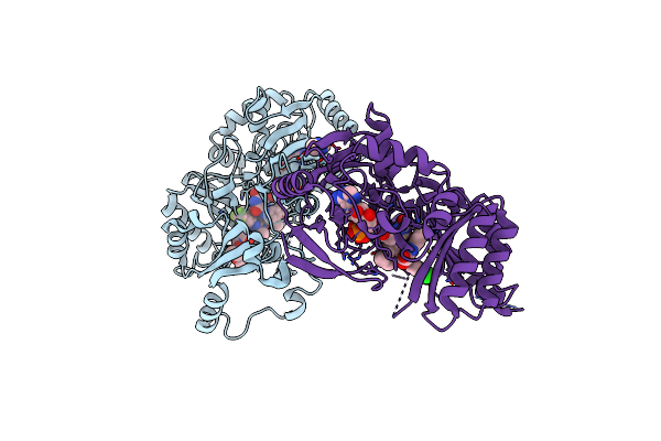
Structures Of Human Ghrelin Receptor-Gi Complexes With Ghrelin And A Synthetic Agonist
Organism: Homo sapiens, Mus musculus
Method: ELECTRON MICROSCOPY Release Date: 2021-12-15 Classification: MEMBRANE PROTEIN Ligands: CLR |
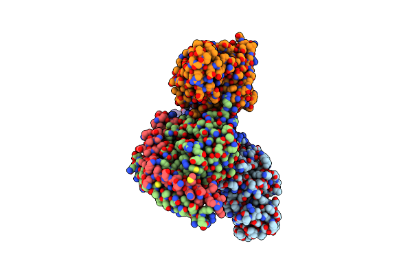 |
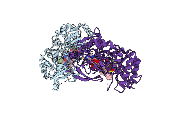
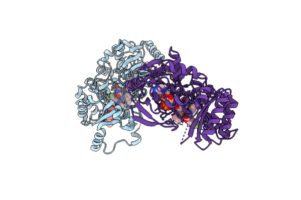
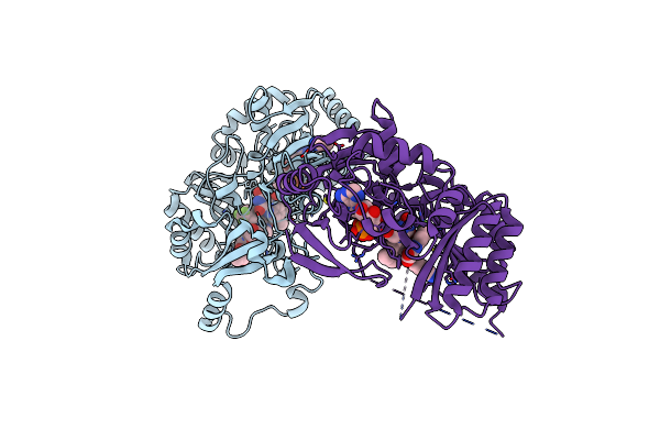
Structures Of Human Ghrelin Receptor-Gi Complexes With Ghrelin And A Synthetic Agonist
Organism: Homo sapiens, Mus musculus
Method: ELECTRON MICROSCOPY Release Date: 2021-12-15 Classification: MEMBRANE PROTEIN Ligands: 1KD, CLR |
 |
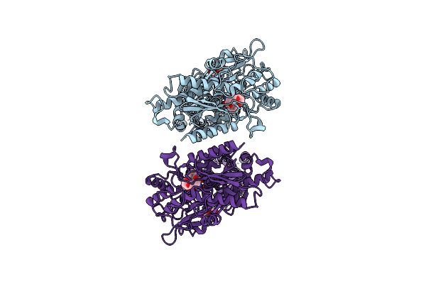
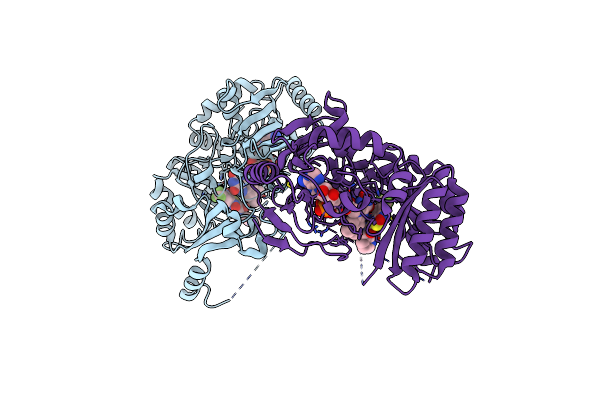
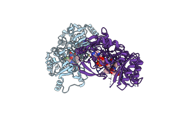
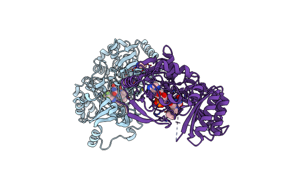
Organism: Homo sapiens
Method: ELECTRON MICROSCOPY Release Date: 2021-09-08 Classification: MEMBRANE PROTEIN Ligands: NAG, QUS, Y01 |
 |
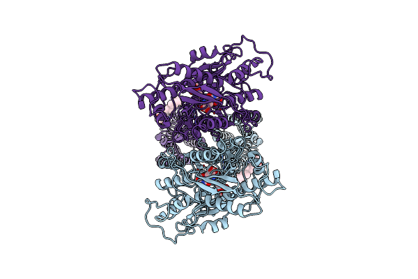
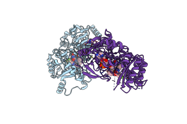
Thermostabilised Full Length Human Mglur5-5M With Orthosteric Antagonist, Ly341495
Organism: Homo sapiens
Method: ELECTRON MICROSCOPY Release Date: 2021-09-08 Classification: MEMBRANE PROTEIN Ligands: NAG, Z99 |
 |
Thermostabilised 7Tm Domain Of Human Mglu5 Receptor Bound To Photoswitchable Ligand Alloswitch-1
Organism: Homo sapiens, Enterobacteria phage t4
Method: X-RAY DIFFRACTION Resolution:2.54 Å Release Date: 2021-09-08 Classification: SIGNALING PROTEIN Ligands: 4YI |
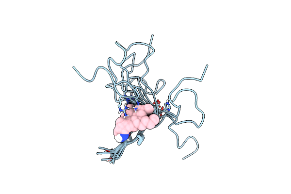 |
Organism: Homo sapiens
Method: SOLUTION NMR Release Date: 2019-07-24 Classification: HORMONE Ligands: FKZ |
 |
Crystal Structure Of M. Tuberculosis Dpre1 In Complex With The Nitro-Benzothiazole 6A
Organism: Mycobacterium tuberculosis
Method: X-RAY DIFFRACTION Resolution:3.00 Å Release Date: 2015-10-28 Classification: OXIDOREDUCTASE/OXIDOREDUCTASE INHIBITOR Ligands: FAD, O95 |
 |
Organism: Mycobacterium tuberculosis
Method: X-RAY DIFFRACTION Resolution:2.56 Å Release Date: 2015-10-21 Classification: oxidoreductase/oxidoreductase inhibitor Ligands: FAD, N77 |
 |
Crystal Structure Of M. Tuberculosis In Complex With A Cbt - Non-Covalent Adduct
Organism: Mycobacterium tuberculosis
Method: X-RAY DIFFRACTION Resolution:2.30 Å Release Date: 2015-10-21 Classification: oxidoreductase/oxidoreductase inhibitor Ligands: FAD, IMD, 2R2 |
 |
Crystal Structure Of The M. Tuberculosis Sulfate Ester Dioxygenase Rv3406 In Complex With Iron.
Organism: Mycobacterium tuberculosis
Method: X-RAY DIFFRACTION Resolution:2.00 Å Release Date: 2014-12-10 Classification: OXIDOREDUCTASE Ligands: NO3, FE |
 |
Crystal Structure Of M. Tuberculosis Dpre1 In Complex With The Non-Covalent Inhibitor Qn127
Organism: Mycobacterium tuberculosis
Method: X-RAY DIFFRACTION Resolution:1.95 Å Release Date: 2014-12-10 Classification: OXIDOREDUCTASE/OXIDOREDUCTASE INHIBITOR Ligands: FAD, Y22, IMD, 2J3 |
 |
Crystal Structure Of M. Tuberculosis Dpre1 In Complex With The Non-Covalent Inhibitor Ty38C
Organism: Mycobacterium tuberculosis
Method: X-RAY DIFFRACTION Resolution:2.49 Å Release Date: 2014-12-10 Classification: OXIDOREDUCTASE/OXIDOREDUCTASE INHIBITOR Ligands: FAD, 38C |
 |
Crystal Structure Of M. Tuberculosis Dpre1 In Complex With The Non-Covalent Inhibitor Ty36C
Organism: Mycobacterium tuberculosis
Method: X-RAY DIFFRACTION Resolution:2.02 Å Release Date: 2014-12-10 Classification: OXIDOREDUCTASE/OXIDOREDUCTASE INHIBITOR Ligands: FAD, 36C, IMD |
 |
Crystal Structure Of M. Tuberculosis Dpre1 In Complex With The Non-Covalent Inhibitor Qn114
Organism: Mycobacterium tuberculosis
Method: X-RAY DIFFRACTION Resolution:2.09 Å Release Date: 2014-12-10 Classification: OXIDOREDUCTASE/OXIDOREDUCTASE INHIBITOR Ligands: FAD, R58, 2J3, IMD |
 |
Crystal Structure Of M. Tuberculosis Dpre1 In Complex With The Non-Covalent Inhibitor Qn118
Organism: Mycobacterium tuberculosis
Method: X-RAY DIFFRACTION Resolution:1.79 Å Release Date: 2014-12-10 Classification: OXIDOREDUCTASE/OXIDOREDUCTASE INHIBITOR Ligands: FAD, R57, IMD |
 |
Crystal Structure Of M. Tuberculosis Dpre1 In Complex With The Non-Covalent Inhibitor Qn127
Organism: Mycobacterium tuberculosis
Method: X-RAY DIFFRACTION Resolution:2.20 Å Release Date: 2014-12-10 Classification: OXIDOREDUCTASE/OXIDOREDUCTASE INHIBITOR Ligands: FAD, RG2, 2J3, IMD |
 |
Crystal Structure Of M. Tuberculosis Dpre1 In Complex With The Non-Covalent Inhibitor Qn129
Organism: Mycobacterium tuberculosis
Method: X-RAY DIFFRACTION Resolution:2.55 Å Release Date: 2014-12-10 Classification: OXIDOREDUCTASE/OXIDOREDUCTASE INHIBITOR Ligands: FAD, R26, IMD, 2J3 |
 |
Crystal Structure Of M. Tuberculosis Dpre1 In Complex With The Non-Covalent Inhibitor Ty21C
Organism: Mycobacterium tuberculosis
Method: X-RAY DIFFRACTION Resolution:2.01 Å Release Date: 2014-12-10 Classification: OXIDOREDUCTASE/OXIDOREDUCTASE INHIBITOR Ligands: FAD, 38C, IMD |
 |
Organism: Mycobacterium tuberculosis
Method: X-RAY DIFFRACTION Resolution:1.88 Å Release Date: 2014-02-19 Classification: OXIDOREDUCTASE/OXIDOREDUCTASE INHIBITOR Ligands: 26J, FAD, GOL, IMD |
 |
Organism: Mycobacterium tuberculosis (strain atcc 25618 / h37rv)
Method: X-RAY DIFFRACTION Resolution:2.25 Å Release Date: 2013-12-04 Classification: OXIDOREDUCTASE Ligands: PYW |