
Search Count: 13
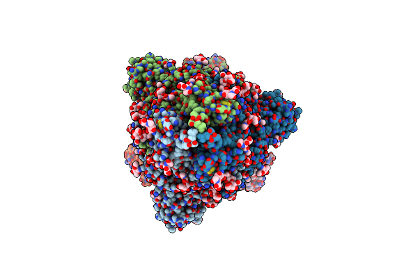 |
Structure Of The Sars-Cov-2 Spike Trimer With All Rbds Down In Complex With The Fab Fragment Of Human Neutralizing Antibody Clone 6
Organism: Severe acute respiratory syndrome coronavirus 2, Homo sapiens
Method: ELECTRON MICROSCOPY Release Date: 2022-03-16 Classification: VIRAL PROTEIN/Immune System Ligands: NAG |
 |
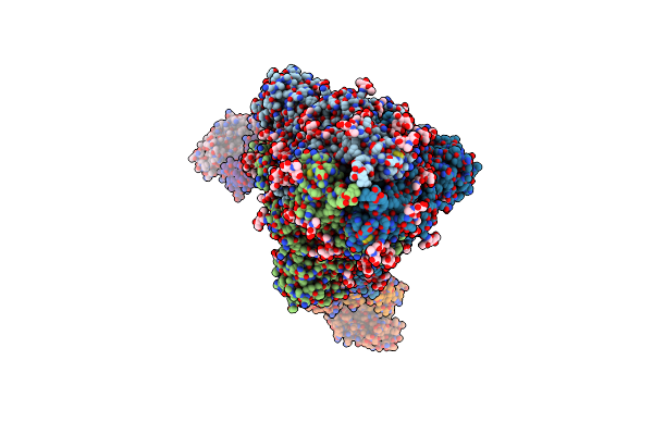
Structure Of The Sars-Cov-2 Spike Trimer With Two Rbds Down In Complex With The Fab Fragment Of Human Neutralizing Antibody Clone 6
Organism: Homo sapiens, Severe acute respiratory syndrome coronavirus 2
Method: ELECTRON MICROSCOPY Release Date: 2022-03-16 Classification: VIRAL PROTEIN/Immune System Ligands: NAG |
 |
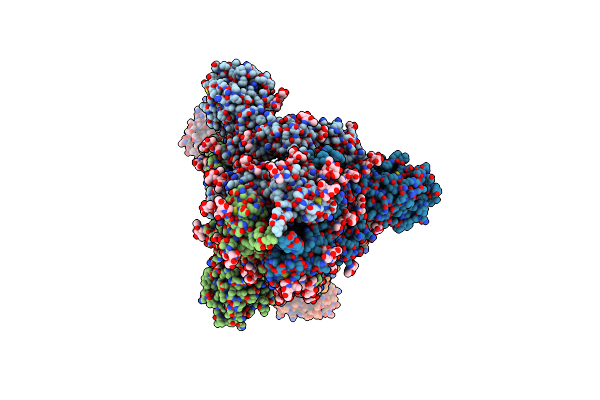
Structure Of The Sars-Cov-2 Spike Trimer With One Rbd Down In Complex With The Fab Fragment Of Human Neutralizing Antibody Clone 6
Organism: Severe acute respiratory syndrome coronavirus 2, Homo sapiens
Method: ELECTRON MICROSCOPY Release Date: 2022-03-16 Classification: VIRAL PROTEIN/Immune System Ligands: NAG |
 |
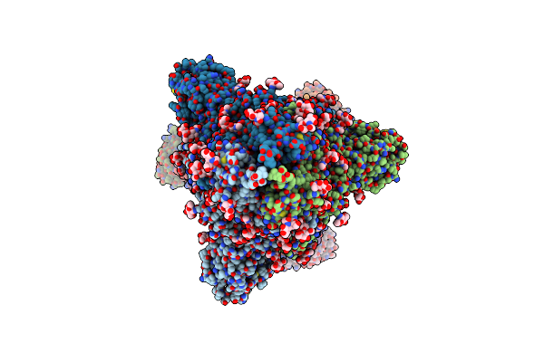
Structure Of The Sars-Cov-2 Spike Trimer With One Rbd Down In Complex With The Fab Fragment Of Human Neutralizing Antibody Clone 2
Organism: Homo sapiens, Severe acute respiratory syndrome coronavirus 2
Method: ELECTRON MICROSCOPY Release Date: 2022-03-16 Classification: VIRAL PROTEIN/Immune System Ligands: NAG |
 |
Structure Of The Sars-Cov-2 Spike Trimer With Three Rbds Up In Complex With The Fab Fragment Of Human Neutralizing Antibody Clone 2
Organism: Severe acute respiratory syndrome coronavirus 2, Homo sapiens
Method: ELECTRON MICROSCOPY Release Date: 2022-03-16 Classification: VIRAL PROTEIN/Immune System Ligands: NAG |
 |
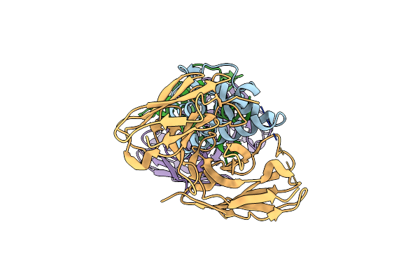
Crystal Structure Of Human Cd81 Large Extracellular Loop In Complex With Single Chain Fv Fragment 4
Organism: Homo sapiens, Mus musculus
Method: X-RAY DIFFRACTION Resolution:2.82 Å Release Date: 2018-05-30 Classification: CELL ADHESION |
 |
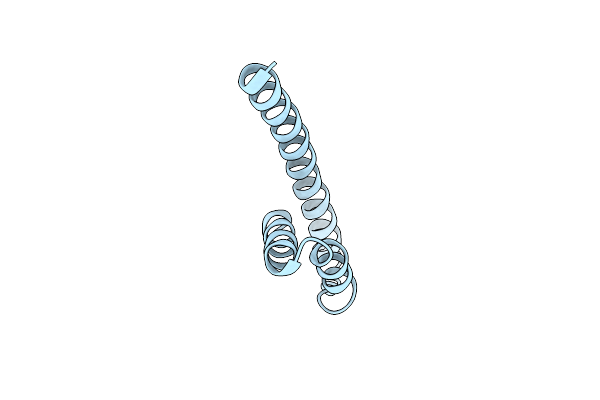
Crystal Structure Of Human Cd81 Large Extracellular Loop In Complex With Single Chain Fv Fragment 5
Organism: Homo sapiens, Mus musculus
Method: X-RAY DIFFRACTION Resolution:2.15 Å Release Date: 2018-05-30 Classification: CELL ADHESION |
 |
Crystal Structure Of Human Cd81 Large Extracellular Loop In Complex With Single Chain Fv Fragment 10
Organism: Homo sapiens, Mus musculus
Method: X-RAY DIFFRACTION Resolution:2.65 Å Release Date: 2018-05-30 Classification: CELL ADHESION |
 |
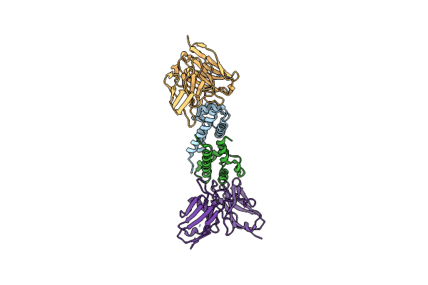
Crystal Structure Of Kit D4D5 Fragment In Complex With Anti-Kit Antibody Fab19
Organism: Homo sapiens
Method: X-RAY DIFFRACTION Resolution:2.40 Å Release Date: 2013-10-16 Classification: IMMUNE SYSTEM Ligands: NAG |
 |
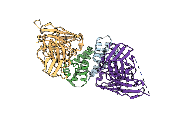
Crystal Structure Of Kit D4D5 Fragment In Complex With Anti-Kit Antibodies Fab79D
Organism: Mus musculus, Homo sapiens
Method: X-RAY DIFFRACTION Resolution:2.70 Å Release Date: 2013-10-16 Classification: IMMUNE SYSTEM Ligands: NAG |
 |
Crystal Structure Of Intracellular Chorismate Mutase From Mycobacterium Tuberculosis
Organism: Mycobacterium tuberculosis
Method: X-RAY DIFFRACTION Resolution:2.00 Å Release Date: 2007-06-26 Classification: ISOMERASE |
 |
Organism: Yersinia pestis biovar microtus str. 91001
Method: X-RAY DIFFRACTION Resolution:2.10 Å Release Date: 2007-04-03 Classification: ISOMERASE Ligands: SO4, CIT |
 |
Organism: Mycobacterium tuberculosis
Method: X-RAY DIFFRACTION Resolution:1.70 Å Release Date: 2005-12-13 Classification: ISOMERASE |